Zinc »
PDB 3lju-3lt9 »
3lk8 »
Zinc in PDB 3lk8: Crystal Structure of the Catalytic Domain of Human MMP12 Complexed with the Inhibitor Paramethoxy-Sulfonyl-Glycine Hydroxamate
Enzymatic activity of Crystal Structure of the Catalytic Domain of Human MMP12 Complexed with the Inhibitor Paramethoxy-Sulfonyl-Glycine Hydroxamate
All present enzymatic activity of Crystal Structure of the Catalytic Domain of Human MMP12 Complexed with the Inhibitor Paramethoxy-Sulfonyl-Glycine Hydroxamate:
3.4.24.65;
3.4.24.65;
Protein crystallography data
The structure of Crystal Structure of the Catalytic Domain of Human MMP12 Complexed with the Inhibitor Paramethoxy-Sulfonyl-Glycine Hydroxamate, PDB code: 3lk8
was solved by
V.Calderone,
with X-Ray Crystallography technique. A brief refinement statistics is given in the table below:
| Resolution Low / High (Å) | 25.81 / 1.80 |
| Space group | C 1 2 1 |
| Cell size a, b, c (Å), α, β, γ (°) | 51.708, 60.251, 54.144, 90.00, 115.16, 90.00 |
| R / Rfree (%) | 16.8 / 20 |
Other elements in 3lk8:
The structure of Crystal Structure of the Catalytic Domain of Human MMP12 Complexed with the Inhibitor Paramethoxy-Sulfonyl-Glycine Hydroxamate also contains other interesting chemical elements:
| Calcium | (Ca) | 3 atoms |
Zinc Binding Sites:
The binding sites of Zinc atom in the Crystal Structure of the Catalytic Domain of Human MMP12 Complexed with the Inhibitor Paramethoxy-Sulfonyl-Glycine Hydroxamate
(pdb code 3lk8). This binding sites where shown within
5.0 Angstroms radius around Zinc atom.
In total 2 binding sites of Zinc where determined in the Crystal Structure of the Catalytic Domain of Human MMP12 Complexed with the Inhibitor Paramethoxy-Sulfonyl-Glycine Hydroxamate, PDB code: 3lk8:
Jump to Zinc binding site number: 1; 2;
In total 2 binding sites of Zinc where determined in the Crystal Structure of the Catalytic Domain of Human MMP12 Complexed with the Inhibitor Paramethoxy-Sulfonyl-Glycine Hydroxamate, PDB code: 3lk8:
Jump to Zinc binding site number: 1; 2;
Zinc binding site 1 out of 2 in 3lk8
Go back to
Zinc binding site 1 out
of 2 in the Crystal Structure of the Catalytic Domain of Human MMP12 Complexed with the Inhibitor Paramethoxy-Sulfonyl-Glycine Hydroxamate
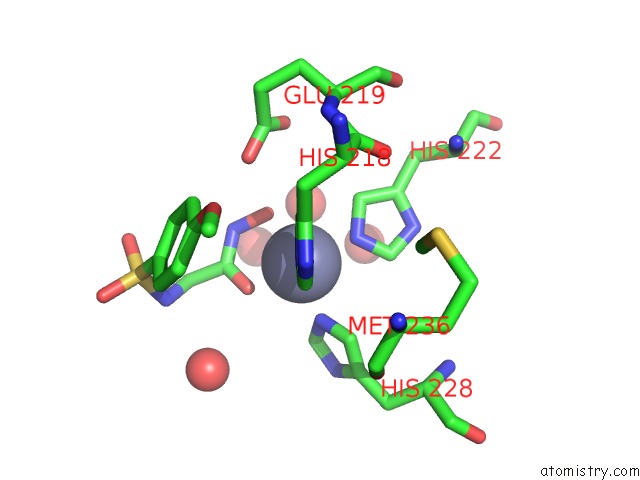
Mono view
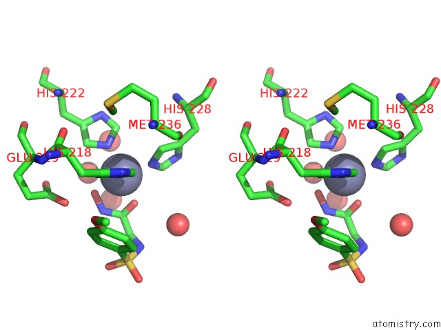
Stereo pair view
Mono view
Stereo pair view
A full contact list of Zinc with other atoms in the Zn binding
site number 1 of Crystal Structure of the Catalytic Domain of Human MMP12 Complexed with the Inhibitor Paramethoxy-Sulfonyl-Glycine Hydroxamate within 5.0Å range:
|
Zinc binding site 2 out of 2 in 3lk8
Go back to
Zinc binding site 2 out
of 2 in the Crystal Structure of the Catalytic Domain of Human MMP12 Complexed with the Inhibitor Paramethoxy-Sulfonyl-Glycine Hydroxamate
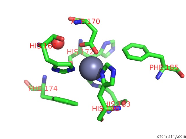
Mono view
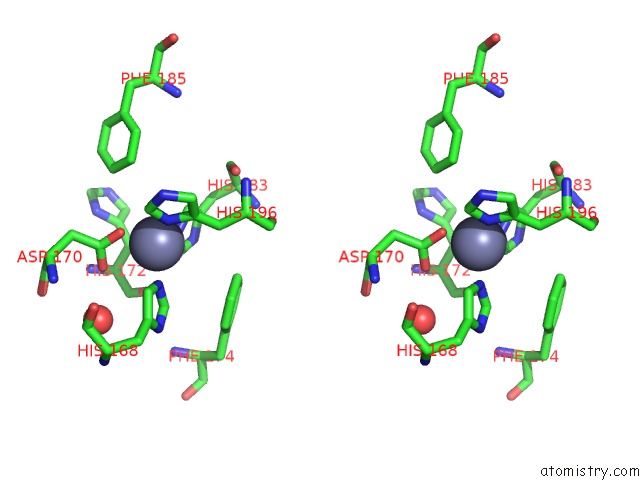
Stereo pair view
Mono view
Stereo pair view
A full contact list of Zinc with other atoms in the Zn binding
site number 2 of Crystal Structure of the Catalytic Domain of Human MMP12 Complexed with the Inhibitor Paramethoxy-Sulfonyl-Glycine Hydroxamate within 5.0Å range:
|
Reference:
I.Bertini,
V.Calderone,
M.Fragai,
A.Giachetti,
M.Loconte,
C.Luchinat,
M.Maletta,
C.Nativi,
K.J.Yeo.
Exploring the Subtleties of Drug-Receptor Interactions: the Case of Matrix Metalloproteinases J.Am.Chem.Soc. V. 129 2466 2007.
ISSN: ISSN 0002-7863
PubMed: 17269766
Page generated: Sat Oct 26 08:41:04 2024
ISSN: ISSN 0002-7863
PubMed: 17269766
Last articles
Zn in 9J0NZn in 9J0O
Zn in 9J0P
Zn in 9FJX
Zn in 9EKB
Zn in 9C0F
Zn in 9CAH
Zn in 9CH0
Zn in 9CH3
Zn in 9CH1