Zinc »
PDB 3k14-3kek »
3kdk »
Zinc in PDB 3kdk: Structure of the C-Terminal Domain of Bacillus Subtilis Mutl Bound to ZN2+
Protein crystallography data
The structure of Structure of the C-Terminal Domain of Bacillus Subtilis Mutl Bound to ZN2+, PDB code: 3kdk
was solved by
A.Guarne,
M.C.Pillon,
with X-Ray Crystallography technique. A brief refinement statistics is given in the table below:
| Resolution Low / High (Å) | 32.75 / 2.26 |
| Space group | P 21 21 21 |
| Cell size a, b, c (Å), α, β, γ (°) | 33.231, 74.608, 182.244, 90.00, 90.00, 90.00 |
| R / Rfree (%) | 21.4 / 26.9 |
Zinc Binding Sites:
The binding sites of Zinc atom in the Structure of the C-Terminal Domain of Bacillus Subtilis Mutl Bound to ZN2+
(pdb code 3kdk). This binding sites where shown within
5.0 Angstroms radius around Zinc atom.
In total 4 binding sites of Zinc where determined in the Structure of the C-Terminal Domain of Bacillus Subtilis Mutl Bound to ZN2+, PDB code: 3kdk:
Jump to Zinc binding site number: 1; 2; 3; 4;
In total 4 binding sites of Zinc where determined in the Structure of the C-Terminal Domain of Bacillus Subtilis Mutl Bound to ZN2+, PDB code: 3kdk:
Jump to Zinc binding site number: 1; 2; 3; 4;
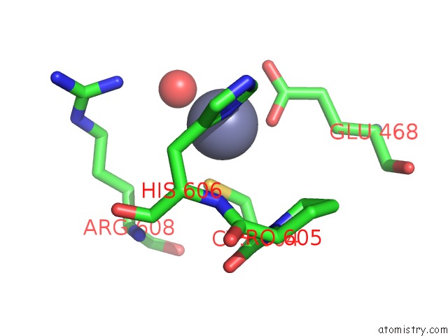
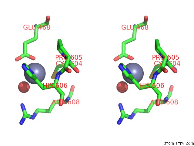
Zinc binding site 1 out of 4 in 3kdk
Go back to
Zinc binding site 1 out
of 4 in the Structure of the C-Terminal Domain of Bacillus Subtilis Mutl Bound to ZN2+
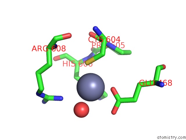
Mono view
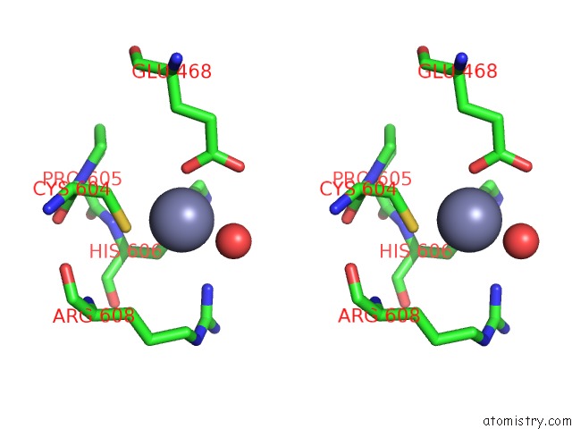
Stereo pair view
Mono view
Stereo pair view
A full contact list of Zinc with other atoms in the Zn binding
site number 1 of Structure of the C-Terminal Domain of Bacillus Subtilis Mutl Bound to ZN2+ within 5.0Å range:
|
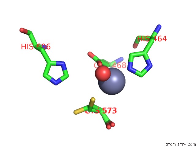
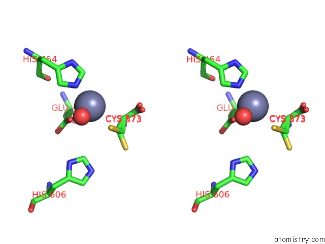
Zinc binding site 2 out of 4 in 3kdk
Go back to
Zinc binding site 2 out
of 4 in the Structure of the C-Terminal Domain of Bacillus Subtilis Mutl Bound to ZN2+
Mono view
Stereo pair view
Mono view
Stereo pair view
A full contact list of Zinc with other atoms in the Zn binding
site number 2 of Structure of the C-Terminal Domain of Bacillus Subtilis Mutl Bound to ZN2+ within 5.0Å range:
|
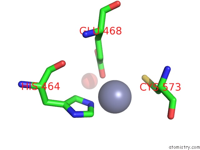
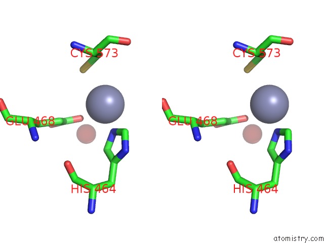
Zinc binding site 3 out of 4 in 3kdk
Go back to
Zinc binding site 3 out
of 4 in the Structure of the C-Terminal Domain of Bacillus Subtilis Mutl Bound to ZN2+
Mono view
Stereo pair view
Mono view
Stereo pair view
A full contact list of Zinc with other atoms in the Zn binding
site number 3 of Structure of the C-Terminal Domain of Bacillus Subtilis Mutl Bound to ZN2+ within 5.0Å range:
|
Zinc binding site 4 out of 4 in 3kdk
Go back to
Zinc binding site 4 out
of 4 in the Structure of the C-Terminal Domain of Bacillus Subtilis Mutl Bound to ZN2+
Mono view
Stereo pair view
Mono view
Stereo pair view
A full contact list of Zinc with other atoms in the Zn binding
site number 4 of Structure of the C-Terminal Domain of Bacillus Subtilis Mutl Bound to ZN2+ within 5.0Å range:
|
Reference:
M.C.Pillon,
J.J.Lorenowicz,
M.Uckelmann,
A.D.Klocko,
R.R.Mitchell,
Y.S.Chung,
P.Modrich,
G.C.Walker,
L.A.Simmons,
P.Friedhoff,
A.Guarne.
Structure of the Endonuclease Domain of Mutl: Unlicensed to Cut. Mol.Cell V. 39 145 2010.
ISSN: ISSN 1097-2765
PubMed: 20603082
DOI: 10.1016/J.MOLCEL.2010.06.027
Page generated: Sat Oct 26 07:45:56 2024
ISSN: ISSN 1097-2765
PubMed: 20603082
DOI: 10.1016/J.MOLCEL.2010.06.027
Last articles
Zn in 9J0NZn in 9J0O
Zn in 9J0P
Zn in 9FJX
Zn in 9EKB
Zn in 9C0F
Zn in 9CAH
Zn in 9CH0
Zn in 9CH3
Zn in 9CH1