Zinc »
PDB 3k14-3kek »
3k7k »
Zinc in PDB 3k7k: Crystal Structure of the Complex Between Carbonic Anhydrase II and Anions
Enzymatic activity of Crystal Structure of the Complex Between Carbonic Anhydrase II and Anions
All present enzymatic activity of Crystal Structure of the Complex Between Carbonic Anhydrase II and Anions:
4.2.1.1;
4.2.1.1;
Protein crystallography data
The structure of Crystal Structure of the Complex Between Carbonic Anhydrase II and Anions, PDB code: 3k7k
was solved by
C.Temperini,
with X-Ray Crystallography technique. A brief refinement statistics is given in the table below:
| Resolution Low / High (Å) | 10.35 / 1.90 |
| Space group | P 1 21 1 |
| Cell size a, b, c (Å), α, β, γ (°) | 42.070, 41.410, 72.230, 90.00, 104.45, 90.00 |
| R / Rfree (%) | 19.4 / 24.8 |
Other elements in 3k7k:
The structure of Crystal Structure of the Complex Between Carbonic Anhydrase II and Anions also contains other interesting chemical elements:
| Mercury | (Hg) | 1 atom |
Zinc Binding Sites:
The binding sites of Zinc atom in the Crystal Structure of the Complex Between Carbonic Anhydrase II and Anions
(pdb code 3k7k). This binding sites where shown within
5.0 Angstroms radius around Zinc atom.
In total only one binding site of Zinc was determined in the Crystal Structure of the Complex Between Carbonic Anhydrase II and Anions, PDB code: 3k7k:
In total only one binding site of Zinc was determined in the Crystal Structure of the Complex Between Carbonic Anhydrase II and Anions, PDB code: 3k7k:
Zinc binding site 1 out of 1 in 3k7k
Go back to
Zinc binding site 1 out
of 1 in the Crystal Structure of the Complex Between Carbonic Anhydrase II and Anions
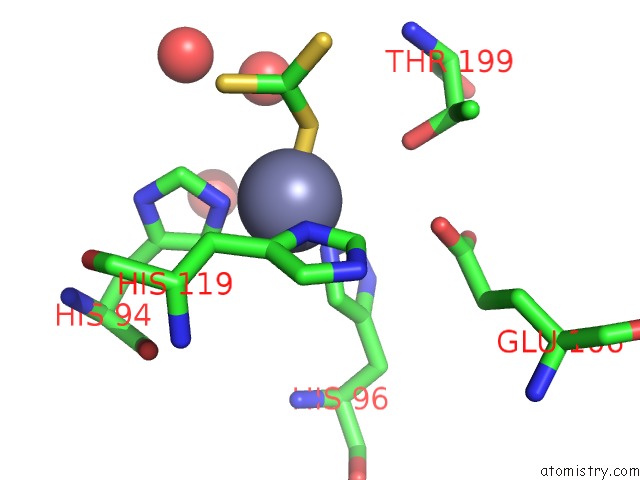
Mono view
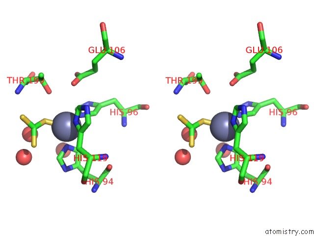
Stereo pair view
Mono view
Stereo pair view
A full contact list of Zinc with other atoms in the Zn binding
site number 1 of Crystal Structure of the Complex Between Carbonic Anhydrase II and Anions within 5.0Å range:
|
Reference:
C.Temperini,
A.Scozzafava,
C.T.Supuran.
Carbonic Anhydrase Inhibitors. X-Ray Crystal Studies of the Carbonic Anhydrase II-Trithiocarbonate Adduct-An Inhibitor Mimicking the Sulfonamide and Urea Binding to the Enzyme Bioorg.Med.Chem.Lett. V. 20 474 2010.
ISSN: ISSN 0960-894X
PubMed: 20005709
DOI: 10.1016/J.BMCL.2009.11.124
Page generated: Sat Oct 26 07:44:15 2024
ISSN: ISSN 0960-894X
PubMed: 20005709
DOI: 10.1016/J.BMCL.2009.11.124
Last articles
Zn in 9J0NZn in 9J0O
Zn in 9J0P
Zn in 9FJX
Zn in 9EKB
Zn in 9C0F
Zn in 9CAH
Zn in 9CH0
Zn in 9CH3
Zn in 9CH1