Zinc »
PDB 3k14-3kek »
3k6i »
Zinc in PDB 3k6i: Crystal Structure of Chicken T-Cadherin EC1
Protein crystallography data
The structure of Crystal Structure of Chicken T-Cadherin EC1, PDB code: 3k6i
was solved by
L.Shapiro,
C.Ciatto,
with X-Ray Crystallography technique. A brief refinement statistics is given in the table below:
| Resolution Low / High (Å) | 18.70 / 1.13 |
| Space group | P 21 21 21 |
| Cell size a, b, c (Å), α, β, γ (°) | 44.153, 44.930, 56.666, 90.00, 90.00, 90.00 |
| R / Rfree (%) | 14.2 / 15.8 |
Zinc Binding Sites:
The binding sites of Zinc atom in the Crystal Structure of Chicken T-Cadherin EC1
(pdb code 3k6i). This binding sites where shown within
5.0 Angstroms radius around Zinc atom.
In total 2 binding sites of Zinc where determined in the Crystal Structure of Chicken T-Cadherin EC1, PDB code: 3k6i:
Jump to Zinc binding site number: 1; 2;
In total 2 binding sites of Zinc where determined in the Crystal Structure of Chicken T-Cadherin EC1, PDB code: 3k6i:
Jump to Zinc binding site number: 1; 2;
Zinc binding site 1 out of 2 in 3k6i
Go back to
Zinc binding site 1 out
of 2 in the Crystal Structure of Chicken T-Cadherin EC1
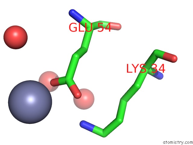
Mono view
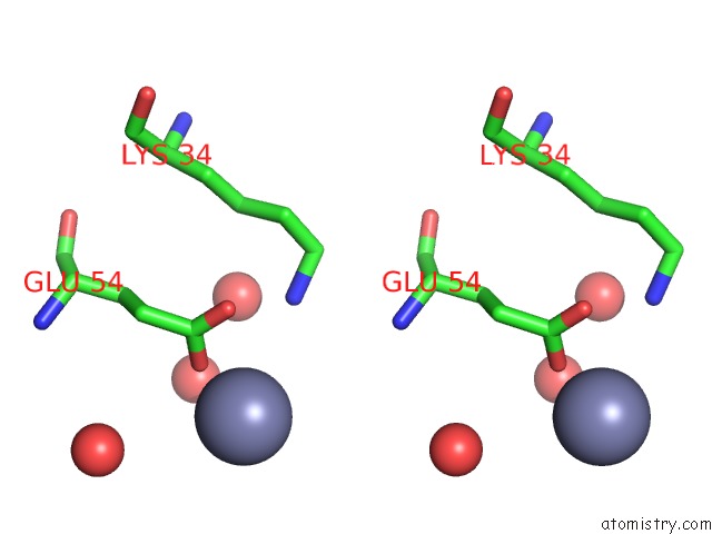
Stereo pair view
Mono view
Stereo pair view
A full contact list of Zinc with other atoms in the Zn binding
site number 1 of Crystal Structure of Chicken T-Cadherin EC1 within 5.0Å range:
|
Zinc binding site 2 out of 2 in 3k6i
Go back to
Zinc binding site 2 out
of 2 in the Crystal Structure of Chicken T-Cadherin EC1
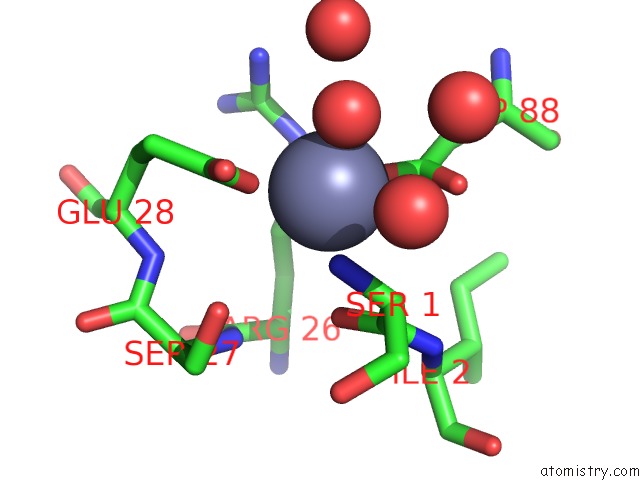
Mono view
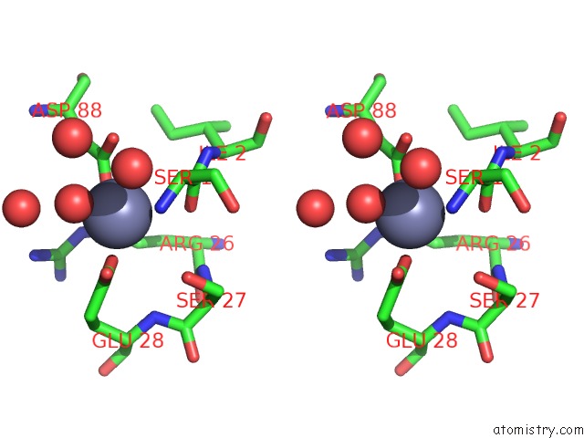
Stereo pair view
Mono view
Stereo pair view
A full contact list of Zinc with other atoms in the Zn binding
site number 2 of Crystal Structure of Chicken T-Cadherin EC1 within 5.0Å range:
|
Reference:
C.Ciatto,
F.Bahna,
N.Zampieri,
H.C.Vansteenhouse,
P.S.Katsamba,
G.Ahlsen,
O.J.Harrison,
J.Brasch,
X.Jin,
S.Posy,
J.Vendome,
B.Ranscht,
T.M.Jessell,
B.Honig,
L.Shapiro.
T-Cadherin Structures Reveal A Novel Adhesive Binding Mechanism Nat.Struct.Mol.Biol. V. 17 339 2010.
ISSN: ISSN 1545-9993
PubMed: 20190755
DOI: 10.1038/NSMB.1781
Page generated: Sat Oct 26 07:41:30 2024
ISSN: ISSN 1545-9993
PubMed: 20190755
DOI: 10.1038/NSMB.1781
Last articles
Zn in 9J0NZn in 9J0O
Zn in 9J0P
Zn in 9FJX
Zn in 9EKB
Zn in 9C0F
Zn in 9CAH
Zn in 9CH0
Zn in 9CH3
Zn in 9CH1