Zinc »
PDB 3k14-3kek »
3k5x »
Zinc in PDB 3k5x: Crystal Structure of Dipeptidase From Streptomics Coelicolor Complexed with Phosphinate Pseudodipeptide L-Ala-D-Asp at 1.4A Resolution.
Protein crystallography data
The structure of Crystal Structure of Dipeptidase From Streptomics Coelicolor Complexed with Phosphinate Pseudodipeptide L-Ala-D-Asp at 1.4A Resolution., PDB code: 3k5x
was solved by
A.A.Fedorov,
E.V.Fedorov,
J.Cummings,
F.M.Raushel,
S.C.Almo,
with X-Ray Crystallography technique. A brief refinement statistics is given in the table below:
| Resolution Low / High (Å) | 24.88 / 1.40 |
| Space group | P 31 2 1 |
| Cell size a, b, c (Å), α, β, γ (°) | 96.910, 96.910, 104.190, 90.00, 90.00, 120.00 |
| R / Rfree (%) | 19.3 / 20.6 |
Zinc Binding Sites:
The binding sites of Zinc atom in the Crystal Structure of Dipeptidase From Streptomics Coelicolor Complexed with Phosphinate Pseudodipeptide L-Ala-D-Asp at 1.4A Resolution.
(pdb code 3k5x). This binding sites where shown within
5.0 Angstroms radius around Zinc atom.
In total 2 binding sites of Zinc where determined in the Crystal Structure of Dipeptidase From Streptomics Coelicolor Complexed with Phosphinate Pseudodipeptide L-Ala-D-Asp at 1.4A Resolution., PDB code: 3k5x:
Jump to Zinc binding site number: 1; 2;
In total 2 binding sites of Zinc where determined in the Crystal Structure of Dipeptidase From Streptomics Coelicolor Complexed with Phosphinate Pseudodipeptide L-Ala-D-Asp at 1.4A Resolution., PDB code: 3k5x:
Jump to Zinc binding site number: 1; 2;
Zinc binding site 1 out of 2 in 3k5x
Go back to
Zinc binding site 1 out
of 2 in the Crystal Structure of Dipeptidase From Streptomics Coelicolor Complexed with Phosphinate Pseudodipeptide L-Ala-D-Asp at 1.4A Resolution.
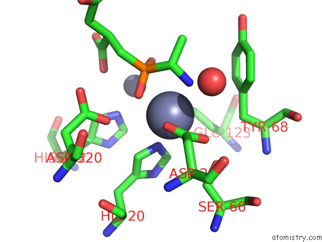
Mono view
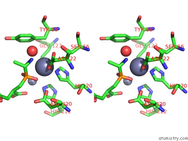
Stereo pair view
Mono view
Stereo pair view
A full contact list of Zinc with other atoms in the Zn binding
site number 1 of Crystal Structure of Dipeptidase From Streptomics Coelicolor Complexed with Phosphinate Pseudodipeptide L-Ala-D-Asp at 1.4A Resolution. within 5.0Å range:
|
Zinc binding site 2 out of 2 in 3k5x
Go back to
Zinc binding site 2 out
of 2 in the Crystal Structure of Dipeptidase From Streptomics Coelicolor Complexed with Phosphinate Pseudodipeptide L-Ala-D-Asp at 1.4A Resolution.
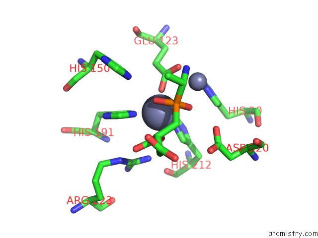
Mono view
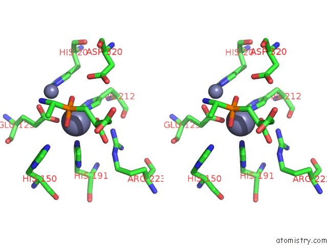
Stereo pair view
Mono view
Stereo pair view
A full contact list of Zinc with other atoms in the Zn binding
site number 2 of Crystal Structure of Dipeptidase From Streptomics Coelicolor Complexed with Phosphinate Pseudodipeptide L-Ala-D-Asp at 1.4A Resolution. within 5.0Å range:
|
Reference:
J.A.Cummings,
T.T.Nguyen,
A.A.Fedorov,
P.Kolb,
C.Xu,
E.V.Fedorov,
B.K.Shoichet,
D.P.Barondeau,
S.C.Almo,
F.M.Raushel.
Structure, Mechanism, and Substrate Profile For SCO3058: the Closest Bacterial Homologue to Human Renal Dipeptidase . Biochemistry V. 49 611 2010.
ISSN: ISSN 0006-2960
PubMed: 20000809
DOI: 10.1021/BI901935Y
Page generated: Sat Oct 26 07:40:30 2024
ISSN: ISSN 0006-2960
PubMed: 20000809
DOI: 10.1021/BI901935Y
Last articles
Zn in 9J0NZn in 9J0O
Zn in 9J0P
Zn in 9FJX
Zn in 9EKB
Zn in 9C0F
Zn in 9CAH
Zn in 9CH0
Zn in 9CH3
Zn in 9CH1