Zinc »
PDB 3jr4-3k0v »
3jt4 »
Zinc in PDB 3jt4: Structure of Neuronal Nitric Oxide Synthase Heme Domain Complexed with N~5~-[(3-(Ethylsulfanyl)Propanimidoyl]-L-Ornithine
Enzymatic activity of Structure of Neuronal Nitric Oxide Synthase Heme Domain Complexed with N~5~-[(3-(Ethylsulfanyl)Propanimidoyl]-L-Ornithine
All present enzymatic activity of Structure of Neuronal Nitric Oxide Synthase Heme Domain Complexed with N~5~-[(3-(Ethylsulfanyl)Propanimidoyl]-L-Ornithine:
1.14.13.39;
1.14.13.39;
Protein crystallography data
The structure of Structure of Neuronal Nitric Oxide Synthase Heme Domain Complexed with N~5~-[(3-(Ethylsulfanyl)Propanimidoyl]-L-Ornithine, PDB code: 3jt4
was solved by
H.Li,
T.L.Poulos,
with X-Ray Crystallography technique. A brief refinement statistics is given in the table below:
| Resolution Low / High (Å) | 37.72 / 1.80 |
| Space group | P 21 21 21 |
| Cell size a, b, c (Å), α, β, γ (°) | 52.026, 111.185, 164.301, 90.00, 90.00, 90.00 |
| R / Rfree (%) | 19.9 / 23.1 |
Other elements in 3jt4:
The structure of Structure of Neuronal Nitric Oxide Synthase Heme Domain Complexed with N~5~-[(3-(Ethylsulfanyl)Propanimidoyl]-L-Ornithine also contains other interesting chemical elements:
| Iron | (Fe) | 2 atoms |
Zinc Binding Sites:
The binding sites of Zinc atom in the Structure of Neuronal Nitric Oxide Synthase Heme Domain Complexed with N~5~-[(3-(Ethylsulfanyl)Propanimidoyl]-L-Ornithine
(pdb code 3jt4). This binding sites where shown within
5.0 Angstroms radius around Zinc atom.
In total only one binding site of Zinc was determined in the Structure of Neuronal Nitric Oxide Synthase Heme Domain Complexed with N~5~-[(3-(Ethylsulfanyl)Propanimidoyl]-L-Ornithine, PDB code: 3jt4:
In total only one binding site of Zinc was determined in the Structure of Neuronal Nitric Oxide Synthase Heme Domain Complexed with N~5~-[(3-(Ethylsulfanyl)Propanimidoyl]-L-Ornithine, PDB code: 3jt4:
Zinc binding site 1 out of 1 in 3jt4
Go back to
Zinc binding site 1 out
of 1 in the Structure of Neuronal Nitric Oxide Synthase Heme Domain Complexed with N~5~-[(3-(Ethylsulfanyl)Propanimidoyl]-L-Ornithine
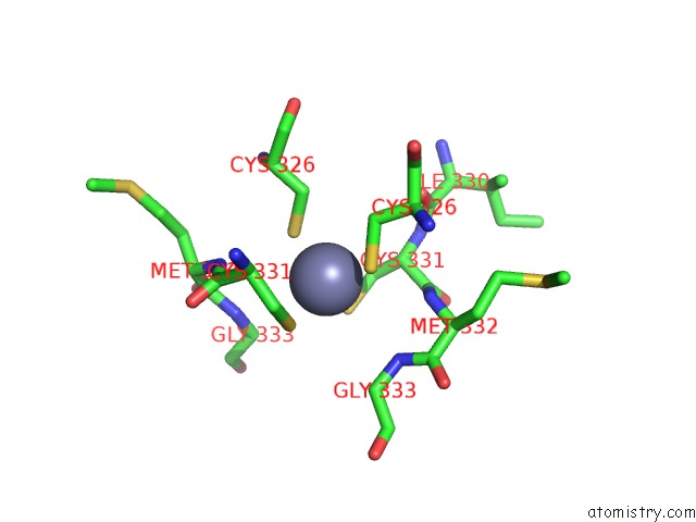
Mono view
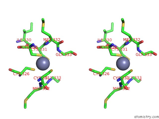
Stereo pair view
Mono view
Stereo pair view
A full contact list of Zinc with other atoms in the Zn binding
site number 1 of Structure of Neuronal Nitric Oxide Synthase Heme Domain Complexed with N~5~-[(3-(Ethylsulfanyl)Propanimidoyl]-L-Ornithine within 5.0Å range:
|
Reference:
J.D.Martell,
H.Li,
T.Doukov,
P.Martasek,
L.J.Roman,
M.Soltis,
T.L.Poulos,
R.B.Silverman.
Heme-Coordinating Inhibitors of Neuronal Nitric Oxide Synthase. Iron-Thioether Coordination Is Stabilized By Hydrophobic Contacts Without Increased Inhibitor Potency. J.Am.Chem.Soc. V. 132 798 2010.
ISSN: ISSN 0002-7863
PubMed: 20014790
DOI: 10.1021/JA908544F
Page generated: Sat Oct 26 07:30:46 2024
ISSN: ISSN 0002-7863
PubMed: 20014790
DOI: 10.1021/JA908544F
Last articles
Zn in 9J0NZn in 9J0O
Zn in 9J0P
Zn in 9FJX
Zn in 9EKB
Zn in 9C0F
Zn in 9CAH
Zn in 9CH0
Zn in 9CH3
Zn in 9CH1