Zinc »
PDB 3iqx-3jr3 »
3iu6 »
Zinc in PDB 3iu6: Crystal Structure of the Sixth Bromodomain of Human Poly-Bromodomain Containing Protein 1 (PB1)
Protein crystallography data
The structure of Crystal Structure of the Sixth Bromodomain of Human Poly-Bromodomain Containing Protein 1 (PB1), PDB code: 3iu6
was solved by
P.Filippakopoulos,
T.Keates,
S.Picaud,
A.C.W.Pike,
E.Ugochukwu,
F.Vondelft,
C.H.Arrowsmith,
A.M.Edwards,
J.Weigelt,
C.Bountra,
S.Knapp,
Structural Genomics Consortium (Sgc),
with X-Ray Crystallography technique. A brief refinement statistics is given in the table below:
| Resolution Low / High (Å) | 33.20 / 1.79 |
| Space group | P 32 2 1 |
| Cell size a, b, c (Å), α, β, γ (°) | 66.400, 66.400, 85.430, 90.00, 90.00, 120.00 |
| R / Rfree (%) | 16.5 / 20.2 |
Zinc Binding Sites:
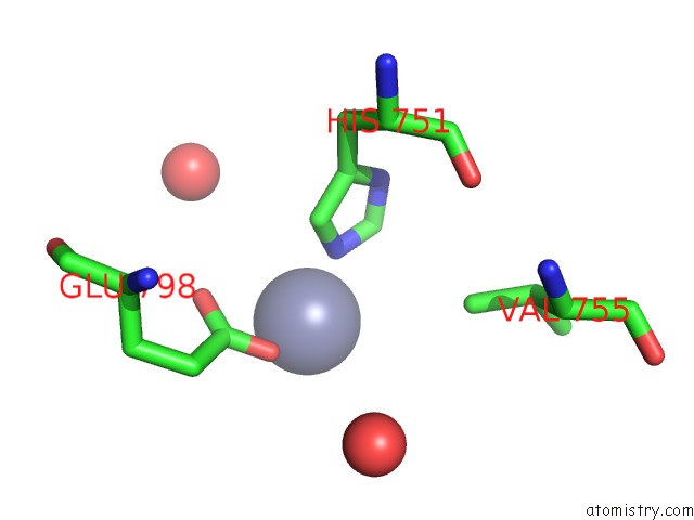
The binding sites of Zinc atom in the Crystal Structure of the Sixth Bromodomain of Human Poly-Bromodomain Containing Protein 1 (PB1)
(pdb code 3iu6). This binding sites where shown within
5.0 Angstroms radius around Zinc atom.
In total only one binding site of Zinc was determined in the Crystal Structure of the Sixth Bromodomain of Human Poly-Bromodomain Containing Protein 1 (PB1), PDB code: 3iu6:
In total only one binding site of Zinc was determined in the Crystal Structure of the Sixth Bromodomain of Human Poly-Bromodomain Containing Protein 1 (PB1), PDB code: 3iu6:
Zinc binding site 1 out of 1 in 3iu6
Go back to
Zinc binding site 1 out
of 1 in the Crystal Structure of the Sixth Bromodomain of Human Poly-Bromodomain Containing Protein 1 (PB1)
Mono view

Stereo pair view
Mono view
Stereo pair view
A full contact list of Zinc with other atoms in the Zn binding
site number 1 of Crystal Structure of the Sixth Bromodomain of Human Poly-Bromodomain Containing Protein 1 (PB1) within 5.0Å range:
|
Reference:
P.Filippakopoulos,
S.Picaud,
M.Mangos,
T.Keates,
J.P.Lambert,
D.Barsyte-Lovejoy,
I.Felletar,
R.Volkmer,
S.Muller,
T.Pawson,
A.C.Gingras,
C.H.Arrowsmith,
S.Knapp.
Histone Recognition and Large-Scale Structural Analysis of the Human Bromodomain Family. Cell(Cambridge,Mass.) V. 149 214 2012.
ISSN: ISSN 0092-8674
PubMed: 22464331
DOI: 10.1016/J.CELL.2012.02.013
Page generated: Sat Oct 26 07:19:59 2024
ISSN: ISSN 0092-8674
PubMed: 22464331
DOI: 10.1016/J.CELL.2012.02.013
Last articles
Zn in 9J0NZn in 9J0O
Zn in 9J0P
Zn in 9FJX
Zn in 9EKB
Zn in 9C0F
Zn in 9CAH
Zn in 9CH0
Zn in 9CH3
Zn in 9CH1