Zinc »
PDB 3iqx-3jr3 »
3ir2 »
Zinc in PDB 3ir2: Crystal Structure of the APOBEC3G Catalytic Domain
Protein crystallography data
The structure of Crystal Structure of the APOBEC3G Catalytic Domain, PDB code: 3ir2
was solved by
S.M.D.Shandilya,
C.A.Schiffer,
with X-Ray Crystallography technique. A brief refinement statistics is given in the table below:
| Resolution Low / High (Å) | 50.00 / 2.25 |
| Space group | P 21 21 21 |
| Cell size a, b, c (Å), α, β, γ (°) | 68.338, 72.532, 97.433, 90.00, 90.00, 90.00 |
| R / Rfree (%) | 16.6 / 20.8 |
Other elements in 3ir2:
The structure of Crystal Structure of the APOBEC3G Catalytic Domain also contains other interesting chemical elements:
| Magnesium | (Mg) | 2 atoms |
| Chlorine | (Cl) | 2 atoms |
Zinc Binding Sites:
The binding sites of Zinc atom in the Crystal Structure of the APOBEC3G Catalytic Domain
(pdb code 3ir2). This binding sites where shown within
5.0 Angstroms radius around Zinc atom.
In total 4 binding sites of Zinc where determined in the Crystal Structure of the APOBEC3G Catalytic Domain, PDB code: 3ir2:
Jump to Zinc binding site number: 1; 2; 3; 4;
In total 4 binding sites of Zinc where determined in the Crystal Structure of the APOBEC3G Catalytic Domain, PDB code: 3ir2:
Jump to Zinc binding site number: 1; 2; 3; 4;
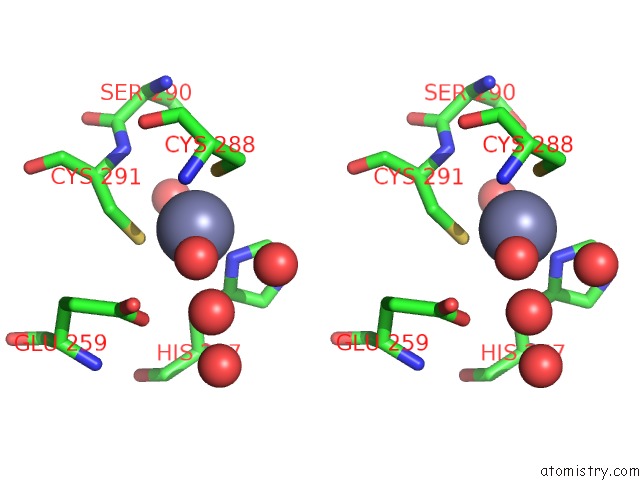
Zinc binding site 1 out of 4 in 3ir2
Go back to
Zinc binding site 1 out
of 4 in the Crystal Structure of the APOBEC3G Catalytic Domain

Mono view

Stereo pair view
Mono view
Stereo pair view
A full contact list of Zinc with other atoms in the Zn binding
site number 1 of Crystal Structure of the APOBEC3G Catalytic Domain within 5.0Å range:
|
Zinc binding site 2 out of 4 in 3ir2
Go back to
Zinc binding site 2 out
of 4 in the Crystal Structure of the APOBEC3G Catalytic Domain

Mono view

Stereo pair view
Mono view
Stereo pair view
A full contact list of Zinc with other atoms in the Zn binding
site number 2 of Crystal Structure of the APOBEC3G Catalytic Domain within 5.0Å range:
|
Zinc binding site 3 out of 4 in 3ir2
Go back to
Zinc binding site 3 out
of 4 in the Crystal Structure of the APOBEC3G Catalytic Domain

Mono view
Stereo pair view
Mono view
Stereo pair view
A full contact list of Zinc with other atoms in the Zn binding
site number 3 of Crystal Structure of the APOBEC3G Catalytic Domain within 5.0Å range:
|
Zinc binding site 4 out of 4 in 3ir2
Go back to
Zinc binding site 4 out
of 4 in the Crystal Structure of the APOBEC3G Catalytic Domain

Mono view

Stereo pair view
Mono view
Stereo pair view
A full contact list of Zinc with other atoms in the Zn binding
site number 4 of Crystal Structure of the APOBEC3G Catalytic Domain within 5.0Å range:
|
Reference:
S.M.Shandilya,
M.N.Nalam,
E.A.Nalivaika,
P.J.Gross,
J.C.Valesano,
K.Shindo,
M.Li,
M.Munson,
W.E.Royer,
E.Harjes,
T.Kono,
H.Matsuo,
R.S.Harris,
M.Somasundaran,
C.A.Schiffer.
Crystal Structure of the APOBEC3G Catalytic Domain Reveals Potential Oligomerization Interfaces. Structure V. 18 28 2010.
ISSN: ISSN 0969-2126
PubMed: 20152150
DOI: 10.1016/J.STR.2009.10.016
Page generated: Sat Oct 26 07:18:01 2024
ISSN: ISSN 0969-2126
PubMed: 20152150
DOI: 10.1016/J.STR.2009.10.016
Last articles
Zn in 9J0NZn in 9J0O
Zn in 9J0P
Zn in 9FJX
Zn in 9EKB
Zn in 9C0F
Zn in 9CAH
Zn in 9CH0
Zn in 9CH3
Zn in 9CH1