Zinc »
PDB 3ifj-3iqx »
3ijf »
Zinc in PDB 3ijf: Crystal Structure of Cytidine Deaminase From Mycobacterium Tuberculosis
Enzymatic activity of Crystal Structure of Cytidine Deaminase From Mycobacterium Tuberculosis
All present enzymatic activity of Crystal Structure of Cytidine Deaminase From Mycobacterium Tuberculosis:
3.5.4.5;
3.5.4.5;
Protein crystallography data
The structure of Crystal Structure of Cytidine Deaminase From Mycobacterium Tuberculosis, PDB code: 3ijf
was solved by
W.F.De Azevedo Jr.,
L.A.Basso,
D.S.Santos,
with X-Ray Crystallography technique. A brief refinement statistics is given in the table below:
| Resolution Low / High (Å) | 18.24 / 1.99 |
| Space group | C 2 2 2 |
| Cell size a, b, c (Å), α, β, γ (°) | 63.705, 75.338, 55.116, 90.00, 90.00, 90.00 |
| R / Rfree (%) | 19 / 24 |
Zinc Binding Sites:
The binding sites of Zinc atom in the Crystal Structure of Cytidine Deaminase From Mycobacterium Tuberculosis
(pdb code 3ijf). This binding sites where shown within
5.0 Angstroms radius around Zinc atom.
In total only one binding site of Zinc was determined in the Crystal Structure of Cytidine Deaminase From Mycobacterium Tuberculosis, PDB code: 3ijf:
In total only one binding site of Zinc was determined in the Crystal Structure of Cytidine Deaminase From Mycobacterium Tuberculosis, PDB code: 3ijf:
Zinc binding site 1 out of 1 in 3ijf
Go back to
Zinc binding site 1 out
of 1 in the Crystal Structure of Cytidine Deaminase From Mycobacterium Tuberculosis
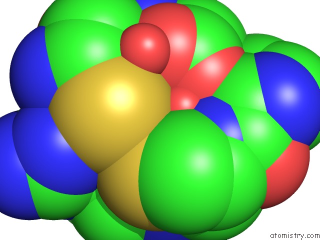
Mono view
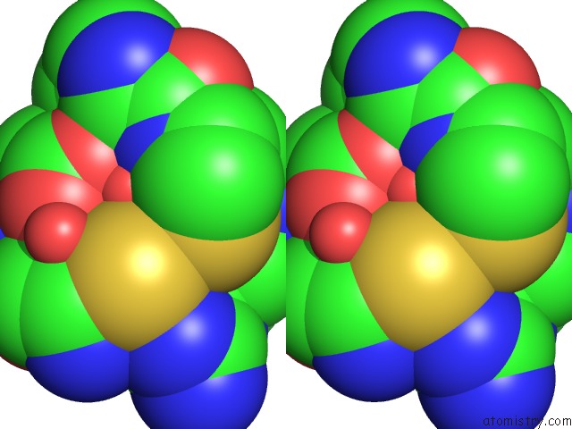
Stereo pair view
Mono view
Stereo pair view
A full contact list of Zinc with other atoms in the Zn binding
site number 1 of Crystal Structure of Cytidine Deaminase From Mycobacterium Tuberculosis within 5.0Å range:
|
Reference:
Z.A.Sanchez-Quitian,
C.Z.Schneider,
R.G.Ducati,
W.F.De Azevedo,
C.Bloch,
L.A.Basso,
D.S.Santos.
Structural and Functional Analyses of Mycobacterium Tuberculosis RV3315C-Encoded Metal-Dependent Homotetrameric Cytidine Deaminase. J.Struct.Biol. V. 169 413 2010.
ISSN: ISSN 1047-8477
PubMed: 20035876
DOI: 10.1016/J.JSB.2009.12.019
Page generated: Wed Aug 20 10:30:56 2025
ISSN: ISSN 1047-8477
PubMed: 20035876
DOI: 10.1016/J.JSB.2009.12.019
Last articles
Zn in 4DZ7Zn in 4DZ9
Zn in 4DYO
Zn in 4DYG
Zn in 4DYK
Zn in 4DY1
Zn in 4DXH
Zn in 4DXX
Zn in 4DXU
Zn in 4DXC