Zinc »
PDB 3ifj-3iqx »
3igp »
Zinc in PDB 3igp: Structure of Inhibitor Binding to Carbonic Anhydrase II
Enzymatic activity of Structure of Inhibitor Binding to Carbonic Anhydrase II
All present enzymatic activity of Structure of Inhibitor Binding to Carbonic Anhydrase II:
4.2.1.1;
4.2.1.1;
Protein crystallography data
The structure of Structure of Inhibitor Binding to Carbonic Anhydrase II, PDB code: 3igp
was solved by
R.Gitto,
S.Agnello,
J.Brynda,
P.Mader,
C.T.Supuran,
A.Chimirri,
with X-Ray Crystallography technique. A brief refinement statistics is given in the table below:
| Resolution Low / High (Å) | 19.95 / 1.65 |
| Space group | P 1 21 1 |
| Cell size a, b, c (Å), α, β, γ (°) | 42.310, 41.270, 72.040, 90.00, 104.22, 90.00 |
| R / Rfree (%) | 15.9 / 19.6 |
Zinc Binding Sites:
The binding sites of Zinc atom in the Structure of Inhibitor Binding to Carbonic Anhydrase II
(pdb code 3igp). This binding sites where shown within
5.0 Angstroms radius around Zinc atom.
In total only one binding site of Zinc was determined in the Structure of Inhibitor Binding to Carbonic Anhydrase II, PDB code: 3igp:
In total only one binding site of Zinc was determined in the Structure of Inhibitor Binding to Carbonic Anhydrase II, PDB code: 3igp:
Zinc binding site 1 out of 1 in 3igp
Go back to
Zinc binding site 1 out
of 1 in the Structure of Inhibitor Binding to Carbonic Anhydrase II
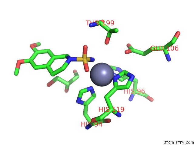
Mono view
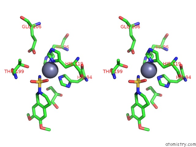
Stereo pair view
Mono view
Stereo pair view
A full contact list of Zinc with other atoms in the Zn binding
site number 1 of Structure of Inhibitor Binding to Carbonic Anhydrase II within 5.0Å range:
|
Reference:
R.Gitto,
S.Agnello,
S.Ferro,
L.De Luca,
D.Vullo,
J.Brynda,
P.Mader,
C.T.Supuran,
A.Chimirri.
Identification of 3,4-Dihydroisoquinoline-2(1H)-Sulfonamides As Potent Carbonic Anhydrase Inhibitors: Synthesis, Biological Evaluation, and Enzyme-Ligand X-Ray Studies. J.Med.Chem. 2010.
ISSN: ISSN 0022-2623
PubMed: 20170095
DOI: 10.1021/JM9014026
Page generated: Wed Aug 20 10:30:16 2025
ISSN: ISSN 0022-2623
PubMed: 20170095
DOI: 10.1021/JM9014026
Last articles
Zn in 4DXXZn in 4DXU
Zn in 4DXC
Zn in 4DXB
Zn in 4DWX
Zn in 4DWV
Zn in 4DWK
Zn in 4DVI
Zn in 4DWC
Zn in 4DV7