Zinc »
PDB 3i7g-3if1 »
3iai »
Zinc in PDB 3iai: Crystal Structure of the Catalytic Domain of the Tumor-Associated Human Carbonic Anhydrase IX
Enzymatic activity of Crystal Structure of the Catalytic Domain of the Tumor-Associated Human Carbonic Anhydrase IX
All present enzymatic activity of Crystal Structure of the Catalytic Domain of the Tumor-Associated Human Carbonic Anhydrase IX:
4.2.1.1;
4.2.1.1;
Protein crystallography data
The structure of Crystal Structure of the Catalytic Domain of the Tumor-Associated Human Carbonic Anhydrase IX, PDB code: 3iai
was solved by
V.Alterio,
A.Di Fiore,
G.De Simone,
with X-Ray Crystallography technique. A brief refinement statistics is given in the table below:
| Resolution Low / High (Å) | 20.00 / 2.20 |
| Space group | P 61 |
| Cell size a, b, c (Å), α, β, γ (°) | 144.180, 144.180, 208.890, 90.00, 90.00, 120.00 |
| R / Rfree (%) | 15.7 / 18.1 |
Zinc Binding Sites:
The binding sites of Zinc atom in the Crystal Structure of the Catalytic Domain of the Tumor-Associated Human Carbonic Anhydrase IX
(pdb code 3iai). This binding sites where shown within
5.0 Angstroms radius around Zinc atom.
In total 4 binding sites of Zinc where determined in the Crystal Structure of the Catalytic Domain of the Tumor-Associated Human Carbonic Anhydrase IX, PDB code: 3iai:
Jump to Zinc binding site number: 1; 2; 3; 4;
In total 4 binding sites of Zinc where determined in the Crystal Structure of the Catalytic Domain of the Tumor-Associated Human Carbonic Anhydrase IX, PDB code: 3iai:
Jump to Zinc binding site number: 1; 2; 3; 4;
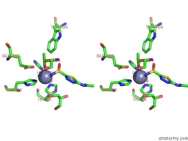
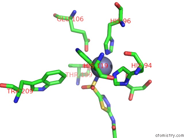
Zinc binding site 1 out of 4 in 3iai
Go back to
Zinc binding site 1 out
of 4 in the Crystal Structure of the Catalytic Domain of the Tumor-Associated Human Carbonic Anhydrase IX
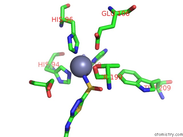
Mono view
Stereo pair view
Mono view
Stereo pair view
A full contact list of Zinc with other atoms in the Zn binding
site number 1 of Crystal Structure of the Catalytic Domain of the Tumor-Associated Human Carbonic Anhydrase IX within 5.0Å range:
|
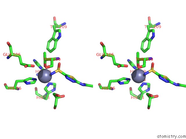
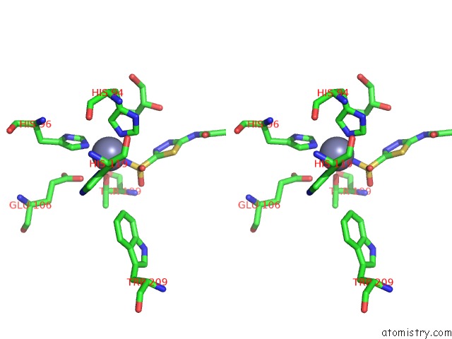
Zinc binding site 2 out of 4 in 3iai
Go back to
Zinc binding site 2 out
of 4 in the Crystal Structure of the Catalytic Domain of the Tumor-Associated Human Carbonic Anhydrase IX
Mono view

Stereo pair view
Mono view
Stereo pair view
A full contact list of Zinc with other atoms in the Zn binding
site number 2 of Crystal Structure of the Catalytic Domain of the Tumor-Associated Human Carbonic Anhydrase IX within 5.0Å range:
|
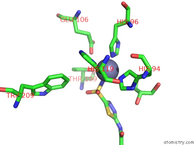
Zinc binding site 3 out of 4 in 3iai
Go back to
Zinc binding site 3 out
of 4 in the Crystal Structure of the Catalytic Domain of the Tumor-Associated Human Carbonic Anhydrase IX
Mono view
Stereo pair view
Mono view
Stereo pair view
A full contact list of Zinc with other atoms in the Zn binding
site number 3 of Crystal Structure of the Catalytic Domain of the Tumor-Associated Human Carbonic Anhydrase IX within 5.0Å range:
|
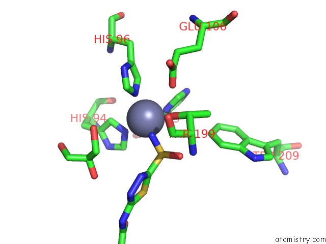
Zinc binding site 4 out of 4 in 3iai
Go back to
Zinc binding site 4 out
of 4 in the Crystal Structure of the Catalytic Domain of the Tumor-Associated Human Carbonic Anhydrase IX
Mono view
Stereo pair view
Mono view
Stereo pair view
A full contact list of Zinc with other atoms in the Zn binding
site number 4 of Crystal Structure of the Catalytic Domain of the Tumor-Associated Human Carbonic Anhydrase IX within 5.0Å range:
|
Reference:
V.Alterio,
M.Hilvo,
A.Di Fiore,
C.T.Supuran,
P.Pan,
S.Parkkila,
A.Scaloni,
J.Pastorek,
S.Pastorekova,
C.Pedone,
A.Scozzafava,
S.M.Monti,
G.De Simone.
Crystal Structure of the Catalytic Domain of the Tumor-Associated Human Carbonic Anhydrase IX. Proc.Natl.Acad.Sci.Usa V. 106 16233 2009.
ISSN: ISSN 0027-8424
PubMed: 19805286
DOI: 10.1073/PNAS.0908301106
Page generated: Sat Oct 26 06:52:25 2024
ISSN: ISSN 0027-8424
PubMed: 19805286
DOI: 10.1073/PNAS.0908301106
Last articles
Zn in 9J0NZn in 9J0O
Zn in 9J0P
Zn in 9FJX
Zn in 9EKB
Zn in 9C0F
Zn in 9CAH
Zn in 9CH0
Zn in 9CH3
Zn in 9CH1