Zinc »
PDB 3h69-3hjw »
3hci »
Zinc in PDB 3hci: Structure of Msrb From Xanthomonas Campestris (Complex-Like Form)
Enzymatic activity of Structure of Msrb From Xanthomonas Campestris (Complex-Like Form)
All present enzymatic activity of Structure of Msrb From Xanthomonas Campestris (Complex-Like Form):
1.8.4.11;
1.8.4.11;
Protein crystallography data
The structure of Structure of Msrb From Xanthomonas Campestris (Complex-Like Form), PDB code: 3hci
was solved by
F.M.Ranaivoson,
B.Kauffmann,
F.Favier,
with X-Ray Crystallography technique. A brief refinement statistics is given in the table below:
| Resolution Low / High (Å) | 24.25 / 2.59 |
| Space group | P 21 21 21 |
| Cell size a, b, c (Å), α, β, γ (°) | 39.436, 74.203, 122.941, 90.00, 90.00, 90.00 |
| R / Rfree (%) | 19.6 / 24.7 |
Other elements in 3hci:
The structure of Structure of Msrb From Xanthomonas Campestris (Complex-Like Form) also contains other interesting chemical elements:
| Calcium | (Ca) | 1 atom |
Zinc Binding Sites:
The binding sites of Zinc atom in the Structure of Msrb From Xanthomonas Campestris (Complex-Like Form)
(pdb code 3hci). This binding sites where shown within
5.0 Angstroms radius around Zinc atom.
In total 2 binding sites of Zinc where determined in the Structure of Msrb From Xanthomonas Campestris (Complex-Like Form), PDB code: 3hci:
Jump to Zinc binding site number: 1; 2;
In total 2 binding sites of Zinc where determined in the Structure of Msrb From Xanthomonas Campestris (Complex-Like Form), PDB code: 3hci:
Jump to Zinc binding site number: 1; 2;
Zinc binding site 1 out of 2 in 3hci
Go back to
Zinc binding site 1 out
of 2 in the Structure of Msrb From Xanthomonas Campestris (Complex-Like Form)
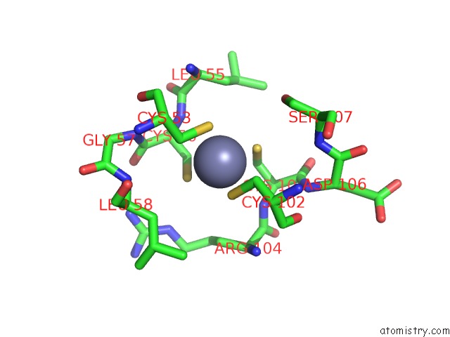
Mono view
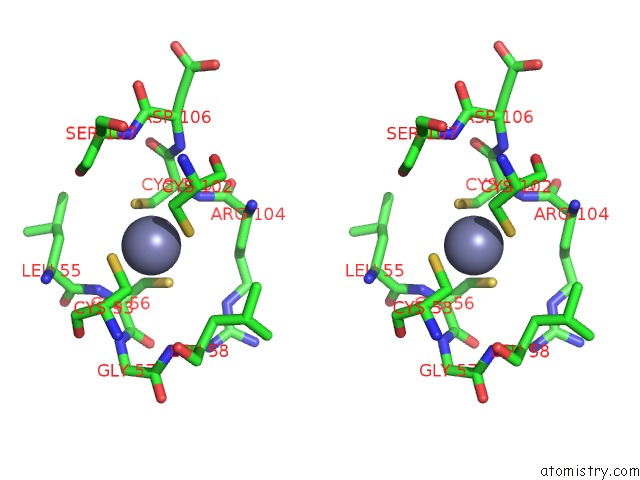
Stereo pair view
Mono view
Stereo pair view
A full contact list of Zinc with other atoms in the Zn binding
site number 1 of Structure of Msrb From Xanthomonas Campestris (Complex-Like Form) within 5.0Å range:
|
Zinc binding site 2 out of 2 in 3hci
Go back to
Zinc binding site 2 out
of 2 in the Structure of Msrb From Xanthomonas Campestris (Complex-Like Form)
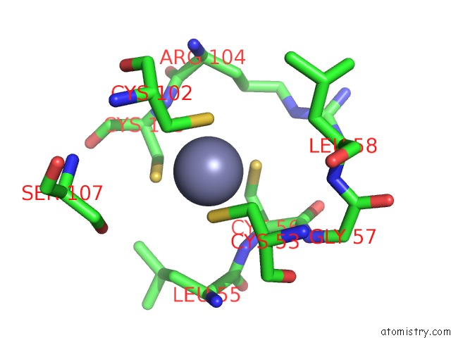
Mono view
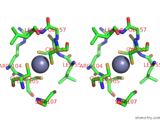
Stereo pair view
Mono view
Stereo pair view
A full contact list of Zinc with other atoms in the Zn binding
site number 2 of Structure of Msrb From Xanthomonas Campestris (Complex-Like Form) within 5.0Å range:
|
Reference:
F.M.Ranaivoson,
F.Neiers,
B.Kauffmann,
S.Boschi-Muller,
G.Branlant,
F.Favier.
Methionine Sulfoxide Reductase B Displays A High Level of Flexibility. J.Mol.Biol. V. 394 83 2009.
ISSN: ISSN 0022-2836
PubMed: 19733575
DOI: 10.1016/J.JMB.2009.08.073
Page generated: Thu Oct 24 14:23:45 2024
ISSN: ISSN 0022-2836
PubMed: 19733575
DOI: 10.1016/J.JMB.2009.08.073
Last articles
Zn in 9J0NZn in 9J0O
Zn in 9J0P
Zn in 9FJX
Zn in 9EKB
Zn in 9C0F
Zn in 9CAH
Zn in 9CH0
Zn in 9CH3
Zn in 9CH1