Zinc »
PDB 3h69-3hjw »
3hbv »
Zinc in PDB 3hbv: Prtc Methionine Mutants: M226A in-House
Protein crystallography data
The structure of Prtc Methionine Mutants: M226A in-House, PDB code: 3hbv
was solved by
A.E.Oberholzer,
M.Bumann,
T.Hege,
S.Russo,
U.Baumann,
with X-Ray Crystallography technique. A brief refinement statistics is given in the table below:
| Resolution Low / High (Å) | 29.35 / 1.95 |
| Space group | P 31 2 1 |
| Cell size a, b, c (Å), α, β, γ (°) | 101.961, 101.961, 123.222, 90.00, 90.00, 120.00 |
| R / Rfree (%) | 15.3 / 16.6 |
Other elements in 3hbv:
The structure of Prtc Methionine Mutants: M226A in-House also contains other interesting chemical elements:
| Chlorine | (Cl) | 4 atoms |
| Calcium | (Ca) | 7 atoms |
Zinc Binding Sites:
The binding sites of Zinc atom in the Prtc Methionine Mutants: M226A in-House
(pdb code 3hbv). This binding sites where shown within
5.0 Angstroms radius around Zinc atom.
In total 2 binding sites of Zinc where determined in the Prtc Methionine Mutants: M226A in-House, PDB code: 3hbv:
Jump to Zinc binding site number: 1; 2;
In total 2 binding sites of Zinc where determined in the Prtc Methionine Mutants: M226A in-House, PDB code: 3hbv:
Jump to Zinc binding site number: 1; 2;
Zinc binding site 1 out of 2 in 3hbv
Go back to
Zinc binding site 1 out
of 2 in the Prtc Methionine Mutants: M226A in-House
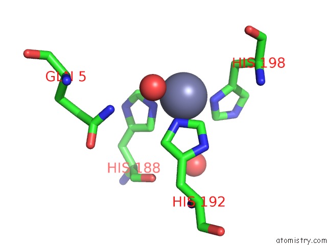
Mono view
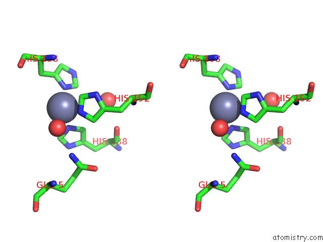
Stereo pair view
Mono view
Stereo pair view
A full contact list of Zinc with other atoms in the Zn binding
site number 1 of Prtc Methionine Mutants: M226A in-House within 5.0Å range:
|
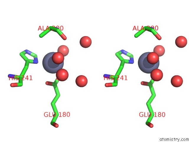
Zinc binding site 2 out of 2 in 3hbv
Go back to
Zinc binding site 2 out
of 2 in the Prtc Methionine Mutants: M226A in-House
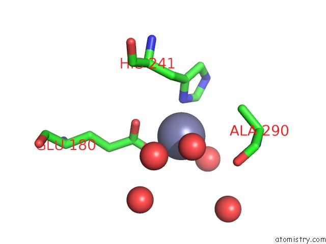
Mono view
Stereo pair view
Mono view
Stereo pair view
A full contact list of Zinc with other atoms in the Zn binding
site number 2 of Prtc Methionine Mutants: M226A in-House within 5.0Å range:
|
Reference:
A.E.Oberholzer,
M.Bumann,
T.Hege,
S.Russo,
U.Baumann.
Metzincin'S Canonical Methionine Is Responsible For the Structural Integrity of the Zinc-Binding Site Biol.Chem. V. 390 875 2009.
ISSN: ISSN 1431-6730
PubMed: 19558324
DOI: 10.1515/BC.2009.100
Page generated: Thu Oct 24 14:22:54 2024
ISSN: ISSN 1431-6730
PubMed: 19558324
DOI: 10.1515/BC.2009.100
Last articles
Zn in 9J0NZn in 9J0O
Zn in 9J0P
Zn in 9FJX
Zn in 9EKB
Zn in 9C0F
Zn in 9CAH
Zn in 9CH0
Zn in 9CH3
Zn in 9CH1