Zinc »
PDB 3h69-3hjw »
3h8v »
Zinc in PDB 3h8v: Human Ubiquitin-Activating Enzyme 5 in Complex with Atp
Protein crystallography data
The structure of Human Ubiquitin-Activating Enzyme 5 in Complex with Atp, PDB code: 3h8v
was solved by
J.R.Walker,
J.P.Bacik,
N.Rastgoo,
J.Weigelt,
C.Bountra,
A.M.Edwards,
C.H.Arrowsmith,
A.Bochkarev,
S.Dhe-Paganon,
Structural Genomicsconsortium (Sgc),
with X-Ray Crystallography technique. A brief refinement statistics is given in the table below:
| Resolution Low / High (Å) | 25.53 / 2.00 |
| Space group | P 32 2 1 |
| Cell size a, b, c (Å), α, β, γ (°) | 77.999, 77.999, 207.005, 90.00, 90.00, 120.00 |
| R / Rfree (%) | 19.1 / 22.4 |
Zinc Binding Sites:
The binding sites of Zinc atom in the Human Ubiquitin-Activating Enzyme 5 in Complex with Atp
(pdb code 3h8v). This binding sites where shown within
5.0 Angstroms radius around Zinc atom.
In total 2 binding sites of Zinc where determined in the Human Ubiquitin-Activating Enzyme 5 in Complex with Atp, PDB code: 3h8v:
Jump to Zinc binding site number: 1; 2;
In total 2 binding sites of Zinc where determined in the Human Ubiquitin-Activating Enzyme 5 in Complex with Atp, PDB code: 3h8v:
Jump to Zinc binding site number: 1; 2;
Zinc binding site 1 out of 2 in 3h8v
Go back to
Zinc binding site 1 out
of 2 in the Human Ubiquitin-Activating Enzyme 5 in Complex with Atp
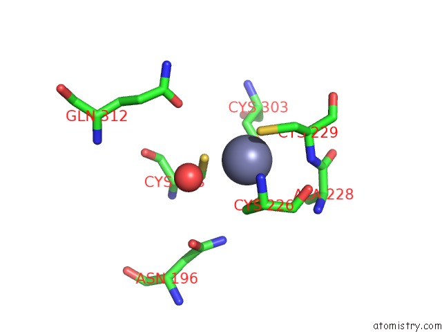
Mono view
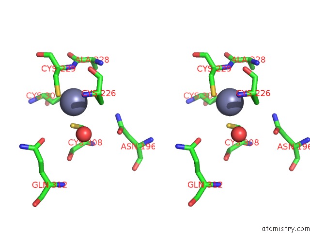
Stereo pair view
Mono view
Stereo pair view
A full contact list of Zinc with other atoms in the Zn binding
site number 1 of Human Ubiquitin-Activating Enzyme 5 in Complex with Atp within 5.0Å range:
|
Zinc binding site 2 out of 2 in 3h8v
Go back to
Zinc binding site 2 out
of 2 in the Human Ubiquitin-Activating Enzyme 5 in Complex with Atp
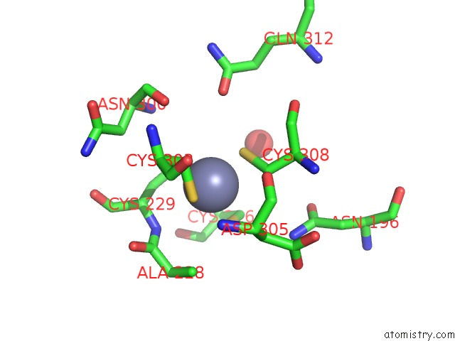
Mono view
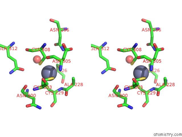
Stereo pair view
Mono view
Stereo pair view
A full contact list of Zinc with other atoms in the Zn binding
site number 2 of Human Ubiquitin-Activating Enzyme 5 in Complex with Atp within 5.0Å range:
|
Reference:
J.P.Bacik,
J.R.Walker,
M.Ali,
A.D.Schimmer,
S.Dhe-Paganon.
Crystal Structure of the Human Ubiquitin-Activating Enzyme 5 (UBA5) Bound to Atp: Mechanistic Insights Into A Minimalistic E1 Enzyme. J.Biol.Chem. V. 285 20273 2010.
ISSN: ISSN 0021-9258
PubMed: 20368332
DOI: 10.1074/JBC.M110.102921
Page generated: Thu Oct 24 14:20:06 2024
ISSN: ISSN 0021-9258
PubMed: 20368332
DOI: 10.1074/JBC.M110.102921
Last articles
Zn in 9J0NZn in 9J0O
Zn in 9J0P
Zn in 9FJX
Zn in 9EKB
Zn in 9C0F
Zn in 9CAH
Zn in 9CH0
Zn in 9CH3
Zn in 9CH1