Zinc »
PDB 3gky-3gtt »
3gn5 »
Zinc in PDB 3gn5: Structure of the E. Coli Protein Mqsa (Ygit/B3021)
Protein crystallography data
The structure of Structure of the E. Coli Protein Mqsa (Ygit/B3021), PDB code: 3gn5
was solved by
B.L.Brown,
J.M.Arruda,
W.Peti,
R.Page,
with X-Ray Crystallography technique. A brief refinement statistics is given in the table below:
| Resolution Low / High (Å) | 50.00 / 2.15 |
| Space group | P 1 21 1 |
| Cell size a, b, c (Å), α, β, γ (°) | 62.054, 30.965, 75.542, 90.00, 106.60, 90.00 |
| R / Rfree (%) | 18.2 / 23.3 |
Zinc Binding Sites:
The binding sites of Zinc atom in the Structure of the E. Coli Protein Mqsa (Ygit/B3021)
(pdb code 3gn5). This binding sites where shown within
5.0 Angstroms radius around Zinc atom.
In total 2 binding sites of Zinc where determined in the Structure of the E. Coli Protein Mqsa (Ygit/B3021), PDB code: 3gn5:
Jump to Zinc binding site number: 1; 2;
In total 2 binding sites of Zinc where determined in the Structure of the E. Coli Protein Mqsa (Ygit/B3021), PDB code: 3gn5:
Jump to Zinc binding site number: 1; 2;
Zinc binding site 1 out of 2 in 3gn5
Go back to
Zinc binding site 1 out
of 2 in the Structure of the E. Coli Protein Mqsa (Ygit/B3021)
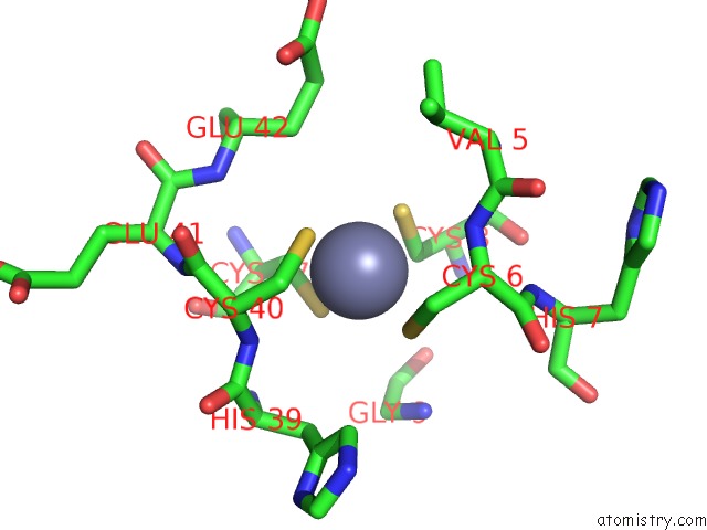
Mono view
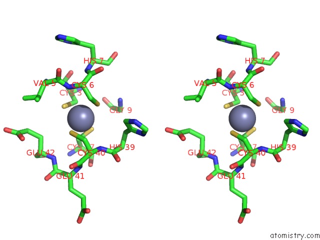
Stereo pair view
Mono view
Stereo pair view
A full contact list of Zinc with other atoms in the Zn binding
site number 1 of Structure of the E. Coli Protein Mqsa (Ygit/B3021) within 5.0Å range:
|
Zinc binding site 2 out of 2 in 3gn5
Go back to
Zinc binding site 2 out
of 2 in the Structure of the E. Coli Protein Mqsa (Ygit/B3021)
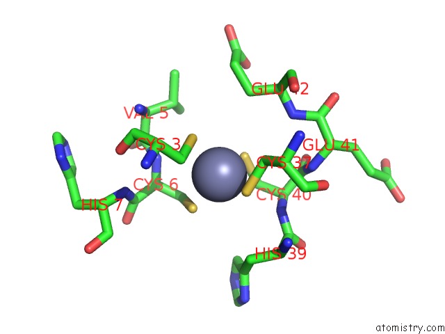
Mono view
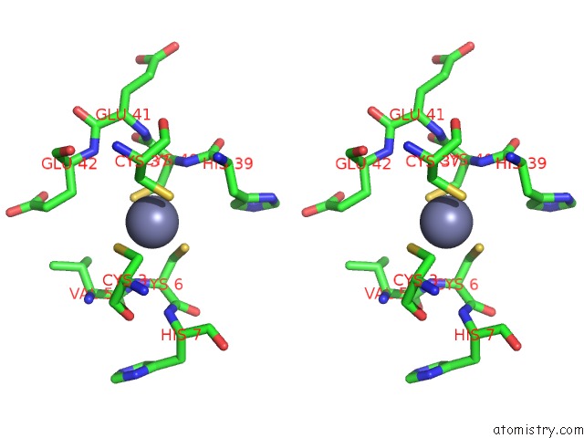
Stereo pair view
Mono view
Stereo pair view
A full contact list of Zinc with other atoms in the Zn binding
site number 2 of Structure of the E. Coli Protein Mqsa (Ygit/B3021) within 5.0Å range:
|
Reference:
B.L.Brown,
S.Grigoriu,
Y.Kim,
J.M.Arruda,
A.Davenport,
T.K.Wood,
W.Peti,
R.Page.
Three Dimensional Structure of the Mqsr:Mqsa Complex: A Novel Ta Pair Comprised of A Toxin Homologous to Rele and An Antitoxin with Unique Properties. Plos Pathog. V. 5 E1000 2009.
ISSN: ISSN 1553-7366
PubMed: 20041169
DOI: 10.1371/JOURNAL.PPAT.1000706
Page generated: Thu Oct 24 13:49:03 2024
ISSN: ISSN 1553-7366
PubMed: 20041169
DOI: 10.1371/JOURNAL.PPAT.1000706
Last articles
Zn in 9J0NZn in 9J0O
Zn in 9J0P
Zn in 9FJX
Zn in 9EKB
Zn in 9C0F
Zn in 9CAH
Zn in 9CH0
Zn in 9CH3
Zn in 9CH1