Zinc »
PDB 3gky-3gtt »
3glr »
Zinc in PDB 3glr: Crystal Structure of Human SIRT3 with Acetyl-Lysine ACECS2 Peptide
Enzymatic activity of Crystal Structure of Human SIRT3 with Acetyl-Lysine ACECS2 Peptide
All present enzymatic activity of Crystal Structure of Human SIRT3 with Acetyl-Lysine ACECS2 Peptide:
6.2.1.1;
6.2.1.1;
Protein crystallography data
The structure of Crystal Structure of Human SIRT3 with Acetyl-Lysine ACECS2 Peptide, PDB code: 3glr
was solved by
L.Jin,
W.Wei,
Y.Jiang,
H.Peng,
J.Cai,
C.Mao,
H.Dai,
J.E.Bemis,
M.R.Jirousek,
J.C.Milne,
C.H.Westphal,
R.B.Perni,
with X-Ray Crystallography technique. A brief refinement statistics is given in the table below:
| Resolution Low / High (Å) | 65.00 / 1.80 |
| Space group | C 2 2 21 |
| Cell size a, b, c (Å), α, β, γ (°) | 78.190, 129.063, 77.899, 90.00, 90.00, 90.00 |
| R / Rfree (%) | 20.3 / 22.6 |
Zinc Binding Sites:
The binding sites of Zinc atom in the Crystal Structure of Human SIRT3 with Acetyl-Lysine ACECS2 Peptide
(pdb code 3glr). This binding sites where shown within
5.0 Angstroms radius around Zinc atom.
In total only one binding site of Zinc was determined in the Crystal Structure of Human SIRT3 with Acetyl-Lysine ACECS2 Peptide, PDB code: 3glr:
In total only one binding site of Zinc was determined in the Crystal Structure of Human SIRT3 with Acetyl-Lysine ACECS2 Peptide, PDB code: 3glr:
Zinc binding site 1 out of 1 in 3glr
Go back to
Zinc binding site 1 out
of 1 in the Crystal Structure of Human SIRT3 with Acetyl-Lysine ACECS2 Peptide
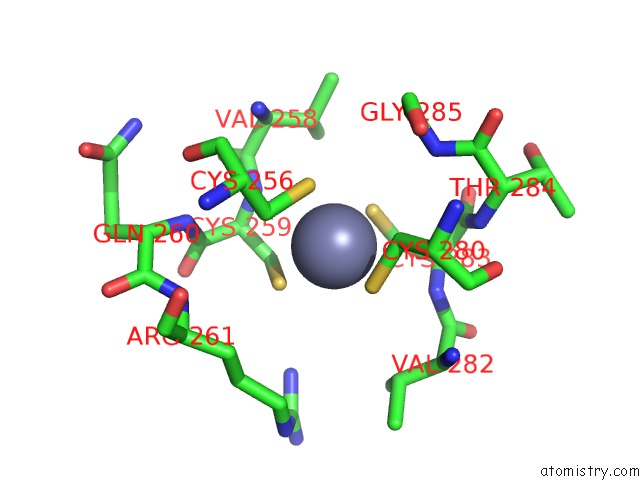
Mono view
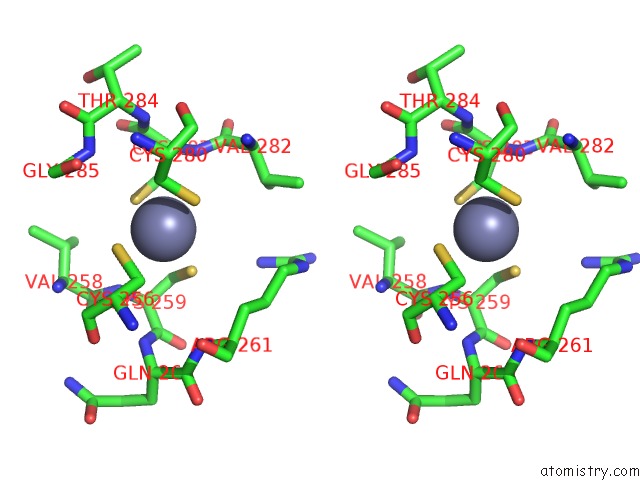
Stereo pair view
Mono view
Stereo pair view
A full contact list of Zinc with other atoms in the Zn binding
site number 1 of Crystal Structure of Human SIRT3 with Acetyl-Lysine ACECS2 Peptide within 5.0Å range:
|
Reference:
L.Jin,
W.Wei,
Y.Jiang,
H.Peng,
J.Cai,
C.Mao,
H.Dai,
W.Choy,
J.E.Bemis,
M.R.Jirousek,
J.C.Milne,
C.H.Westphal,
R.B.Perni.
Crystal Structures of Human SIRT3 Displaying Substrate-Induced Conformational Changes. J.Biol.Chem. V. 284 24394 2009.
ISSN: ISSN 0021-9258
PubMed: 19535340
DOI: 10.1074/JBC.M109.014928
Page generated: Thu Oct 24 13:46:23 2024
ISSN: ISSN 0021-9258
PubMed: 19535340
DOI: 10.1074/JBC.M109.014928
Last articles
Zn in 9J0NZn in 9J0O
Zn in 9J0P
Zn in 9FJX
Zn in 9EKB
Zn in 9C0F
Zn in 9CAH
Zn in 9CH0
Zn in 9CH3
Zn in 9CH1