Zinc »
PDB 3flf-3fuk »
3fua »
Zinc in PDB 3fua: L-Fuculose-1-Phosphate Aldolase Crystal Form K
Enzymatic activity of L-Fuculose-1-Phosphate Aldolase Crystal Form K
All present enzymatic activity of L-Fuculose-1-Phosphate Aldolase Crystal Form K:
4.1.2.17;
4.1.2.17;
Protein crystallography data
The structure of L-Fuculose-1-Phosphate Aldolase Crystal Form K, PDB code: 3fua
was solved by
M.K.Dreyer,
G.E.Schulz,
with X-Ray Crystallography technique. A brief refinement statistics is given in the table below:
| Resolution Low / High (Å) | 10.00 / 2.67 |
| Space group | F 4 3 2 |
| Cell size a, b, c (Å), α, β, γ (°) | 183.000, 183.000, 183.000, 90.00, 90.00, 90.00 |
| R / Rfree (%) | 18.4 / 24.1 |
Other elements in 3fua:
The structure of L-Fuculose-1-Phosphate Aldolase Crystal Form K also contains other interesting chemical elements:
| Chlorine | (Cl) | 1 atom |
Zinc Binding Sites:
The binding sites of Zinc atom in the L-Fuculose-1-Phosphate Aldolase Crystal Form K
(pdb code 3fua). This binding sites where shown within
5.0 Angstroms radius around Zinc atom.
In total only one binding site of Zinc was determined in the L-Fuculose-1-Phosphate Aldolase Crystal Form K, PDB code: 3fua:
In total only one binding site of Zinc was determined in the L-Fuculose-1-Phosphate Aldolase Crystal Form K, PDB code: 3fua:
Zinc binding site 1 out of 1 in 3fua
Go back to
Zinc binding site 1 out
of 1 in the L-Fuculose-1-Phosphate Aldolase Crystal Form K
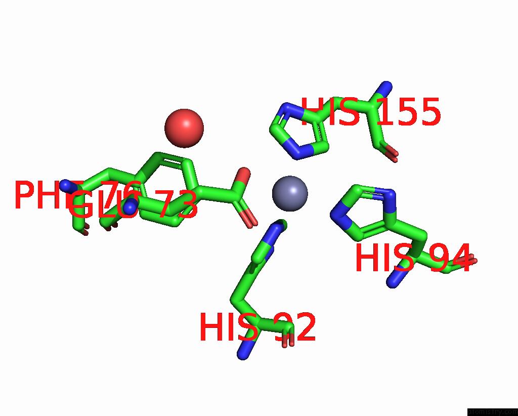
Mono view
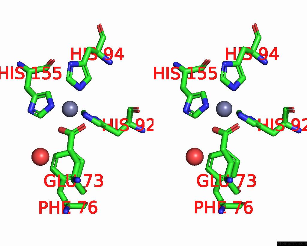
Stereo pair view
Mono view
Stereo pair view
A full contact list of Zinc with other atoms in the Zn binding
site number 1 of L-Fuculose-1-Phosphate Aldolase Crystal Form K within 5.0Å range:
|
Reference:
M.K.Dreyer,
G.E.Schulz.
Catalytic Mechanism of the Metal-Dependent Fuculose Aldolase From Escherichia Coli As Derived From the Structure. J.Mol.Biol. V. 259 458 1996.
ISSN: ISSN 0022-2836
PubMed: 8676381
DOI: 10.1006/JMBI.1996.0332
Page generated: Thu Oct 24 13:25:39 2024
ISSN: ISSN 0022-2836
PubMed: 8676381
DOI: 10.1006/JMBI.1996.0332
Last articles
Zn in 9J0NZn in 9J0O
Zn in 9J0P
Zn in 9FJX
Zn in 9EKB
Zn in 9C0F
Zn in 9CAH
Zn in 9CH0
Zn in 9CH3
Zn in 9CH1