Zinc »
PDB 3ebe-3ekl »
3eft »
Zinc in PDB 3eft: Crystal Structure of the Complex Between Carbonic Anhydrase II and A Spin-Labeled Sulfonamide Incorporating Tempo Moiety
Enzymatic activity of Crystal Structure of the Complex Between Carbonic Anhydrase II and A Spin-Labeled Sulfonamide Incorporating Tempo Moiety
All present enzymatic activity of Crystal Structure of the Complex Between Carbonic Anhydrase II and A Spin-Labeled Sulfonamide Incorporating Tempo Moiety:
4.2.1.1;
4.2.1.1;
Protein crystallography data
The structure of Crystal Structure of the Complex Between Carbonic Anhydrase II and A Spin-Labeled Sulfonamide Incorporating Tempo Moiety, PDB code: 3eft
was solved by
C.Temperini,
A.Cecchi,
A.Scozzafava,
C.T.Supuran,
with X-Ray Crystallography technique. A brief refinement statistics is given in the table below:
| Resolution Low / High (Å) | 11.81 / 1.85 |
| Space group | P 1 21 1 |
| Cell size a, b, c (Å), α, β, γ (°) | 42.130, 41.500, 72.310, 90.00, 104.30, 90.00 |
| R / Rfree (%) | 19.2 / 24 |
Other elements in 3eft:
The structure of Crystal Structure of the Complex Between Carbonic Anhydrase II and A Spin-Labeled Sulfonamide Incorporating Tempo Moiety also contains other interesting chemical elements:
| Mercury | (Hg) | 1 atom |
| Chlorine | (Cl) | 1 atom |
Zinc Binding Sites:
The binding sites of Zinc atom in the Crystal Structure of the Complex Between Carbonic Anhydrase II and A Spin-Labeled Sulfonamide Incorporating Tempo Moiety
(pdb code 3eft). This binding sites where shown within
5.0 Angstroms radius around Zinc atom.
In total only one binding site of Zinc was determined in the Crystal Structure of the Complex Between Carbonic Anhydrase II and A Spin-Labeled Sulfonamide Incorporating Tempo Moiety, PDB code: 3eft:
In total only one binding site of Zinc was determined in the Crystal Structure of the Complex Between Carbonic Anhydrase II and A Spin-Labeled Sulfonamide Incorporating Tempo Moiety, PDB code: 3eft:
Zinc binding site 1 out of 1 in 3eft
Go back to
Zinc binding site 1 out
of 1 in the Crystal Structure of the Complex Between Carbonic Anhydrase II and A Spin-Labeled Sulfonamide Incorporating Tempo Moiety
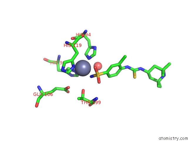
Mono view
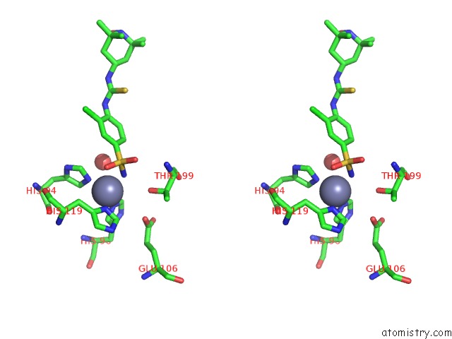
Stereo pair view
Mono view
Stereo pair view
A full contact list of Zinc with other atoms in the Zn binding
site number 1 of Crystal Structure of the Complex Between Carbonic Anhydrase II and A Spin-Labeled Sulfonamide Incorporating Tempo Moiety within 5.0Å range:
|
Reference:
L.Ciani,
A.Cecchi,
C.Temperini,
C.T.Supuran,
S.Ristori.
Dissecting the Inhibition Mechanism of Cytosolic Versus Transmembrane Carbonic Anhydrases By Esr J.Phys.Chem.B V. 113 13998 2009.
ISSN: ISSN 1089-5647
PubMed: 19778001
DOI: 10.1021/JP906593C
Page generated: Thu Oct 24 12:47:46 2024
ISSN: ISSN 1089-5647
PubMed: 19778001
DOI: 10.1021/JP906593C
Last articles
Zn in 9J0NZn in 9J0O
Zn in 9J0P
Zn in 9FJX
Zn in 9EKB
Zn in 9C0F
Zn in 9CAH
Zn in 9CH0
Zn in 9CH3
Zn in 9CH1