Zinc »
PDB 3e2u-3eb9 »
3eah »
Zinc in PDB 3eah: Structure of Inhibited Human Enos Oxygenase Domain
Enzymatic activity of Structure of Inhibited Human Enos Oxygenase Domain
All present enzymatic activity of Structure of Inhibited Human Enos Oxygenase Domain:
1.14.13.39;
1.14.13.39;
Protein crystallography data
The structure of Structure of Inhibited Human Enos Oxygenase Domain, PDB code: 3eah
was solved by
E.D.Garcin,
A.S.Arvai,
R.J.Rosenfeld,
M.D.Kroeger,
B.R.Crane,
G.Andersson,
G.Andrews,
P.J.Hamley,
P.R.Mallinder,
D.J.Nicholls,
S.A.St-Gallay,
A.C.Tinker,
N.P.Gensmantel,
A.Mete,
D.R.Cheshire,
S.Connolly,
D.J.Stuehr,
A.Aberg,
A.V.Wallace,
J.A.Tainer,
E.D.Getzoff,
with X-Ray Crystallography technique. A brief refinement statistics is given in the table below:
| Resolution Low / High (Å) | 25.00 / 2.44 |
| Space group | P 21 21 21 |
| Cell size a, b, c (Å), α, β, γ (°) | 71.090, 90.150, 156.100, 90.00, 90.00, 90.00 |
| R / Rfree (%) | 21.3 / 25.8 |
Other elements in 3eah:
The structure of Structure of Inhibited Human Enos Oxygenase Domain also contains other interesting chemical elements:
| Iron | (Fe) | 2 atoms |
| Chlorine | (Cl) | 2 atoms |
Zinc Binding Sites:
The binding sites of Zinc atom in the Structure of Inhibited Human Enos Oxygenase Domain
(pdb code 3eah). This binding sites where shown within
5.0 Angstroms radius around Zinc atom.
In total only one binding site of Zinc was determined in the Structure of Inhibited Human Enos Oxygenase Domain, PDB code: 3eah:
In total only one binding site of Zinc was determined in the Structure of Inhibited Human Enos Oxygenase Domain, PDB code: 3eah:
Zinc binding site 1 out of 1 in 3eah
Go back to
Zinc binding site 1 out
of 1 in the Structure of Inhibited Human Enos Oxygenase Domain
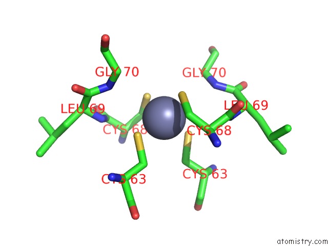
Mono view
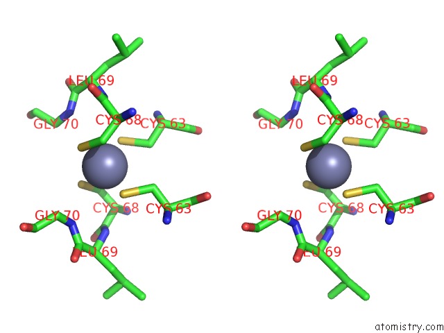
Stereo pair view
Mono view
Stereo pair view
A full contact list of Zinc with other atoms in the Zn binding
site number 1 of Structure of Inhibited Human Enos Oxygenase Domain within 5.0Å range:
|
Reference:
E.D.Garcin,
A.S.Arvai,
R.J.Rosenfeld,
M.D.Kroeger,
B.R.Crane,
G.Andersson,
G.Andrews,
P.J.Hamley,
P.R.Mallinder,
D.J.Nicholls,
S.A.St-Gallay,
A.C.Tinker,
N.P.Gensmantel,
A.Mete,
D.R.Cheshire,
S.Connolly,
D.J.Stuehr,
A.Aberg,
A.V.Wallace,
J.A.Tainer,
E.D.Getzoff.
Anchored Plasticity Opens Doors For Selective Inhibitor Design in Nitric Oxide Synthase. Nat.Chem.Biol. V. 4 700 2008.
ISSN: ISSN 1552-4450
PubMed: 18849972
DOI: 10.1038/NCHEMBIO.115
Page generated: Thu Oct 24 12:41:50 2024
ISSN: ISSN 1552-4450
PubMed: 18849972
DOI: 10.1038/NCHEMBIO.115
Last articles
Zn in 9J0NZn in 9J0O
Zn in 9J0P
Zn in 9FJX
Zn in 9EKB
Zn in 9C0F
Zn in 9CAH
Zn in 9CH0
Zn in 9CH3
Zn in 9CH1