Zinc »
PDB 3e2u-3eb9 »
3ea2 »
Zinc in PDB 3ea2: Crystal Structure of the Myo-Inositol Bound Y247S/Y251S Mutant of Phosphatidylinositol-Specific Phospholipase C From Bacillus Thuringiensis
Enzymatic activity of Crystal Structure of the Myo-Inositol Bound Y247S/Y251S Mutant of Phosphatidylinositol-Specific Phospholipase C From Bacillus Thuringiensis
All present enzymatic activity of Crystal Structure of the Myo-Inositol Bound Y247S/Y251S Mutant of Phosphatidylinositol-Specific Phospholipase C From Bacillus Thuringiensis:
4.6.1.13;
4.6.1.13;
Protein crystallography data
The structure of Crystal Structure of the Myo-Inositol Bound Y247S/Y251S Mutant of Phosphatidylinositol-Specific Phospholipase C From Bacillus Thuringiensis, PDB code: 3ea2
was solved by
X.Shi,
C.Shao,
X.Zhang,
C.Zambonelli,
A.G.Redfied,
J.F.Head,
B.A.Seaton,
M.F.Roberts,
with X-Ray Crystallography technique. A brief refinement statistics is given in the table below:
| Resolution Low / High (Å) | 50.00 / 1.95 |
| Space group | P 21 21 21 |
| Cell size a, b, c (Å), α, β, γ (°) | 59.518, 97.226, 112.727, 90.00, 90.00, 90.00 |
| R / Rfree (%) | 18.9 / 22.6 |
Zinc Binding Sites:
The binding sites of Zinc atom in the Crystal Structure of the Myo-Inositol Bound Y247S/Y251S Mutant of Phosphatidylinositol-Specific Phospholipase C From Bacillus Thuringiensis
(pdb code 3ea2). This binding sites where shown within
5.0 Angstroms radius around Zinc atom.
In total 6 binding sites of Zinc where determined in the Crystal Structure of the Myo-Inositol Bound Y247S/Y251S Mutant of Phosphatidylinositol-Specific Phospholipase C From Bacillus Thuringiensis, PDB code: 3ea2:
Jump to Zinc binding site number: 1; 2; 3; 4; 5; 6;
In total 6 binding sites of Zinc where determined in the Crystal Structure of the Myo-Inositol Bound Y247S/Y251S Mutant of Phosphatidylinositol-Specific Phospholipase C From Bacillus Thuringiensis, PDB code: 3ea2:
Jump to Zinc binding site number: 1; 2; 3; 4; 5; 6;
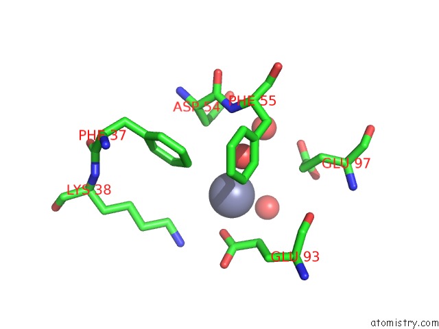
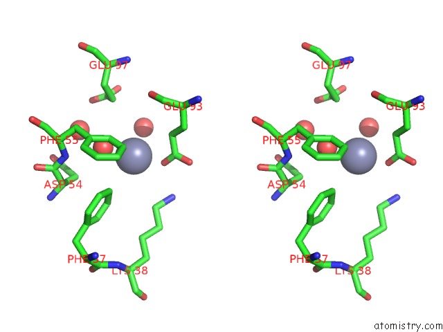
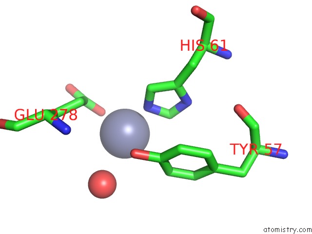
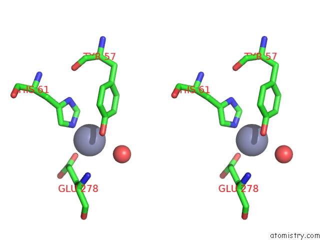
Zinc binding site 1 out of 6 in 3ea2
Go back to
Zinc binding site 1 out
of 6 in the Crystal Structure of the Myo-Inositol Bound Y247S/Y251S Mutant of Phosphatidylinositol-Specific Phospholipase C From Bacillus Thuringiensis
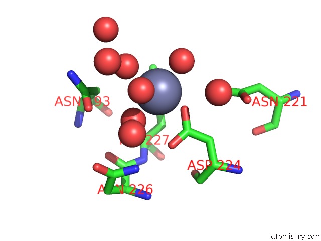
Mono view
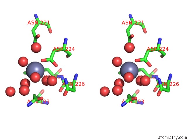
Stereo pair view
Mono view
Stereo pair view
A full contact list of Zinc with other atoms in the Zn binding
site number 1 of Crystal Structure of the Myo-Inositol Bound Y247S/Y251S Mutant of Phosphatidylinositol-Specific Phospholipase C From Bacillus Thuringiensis within 5.0Å range:
|
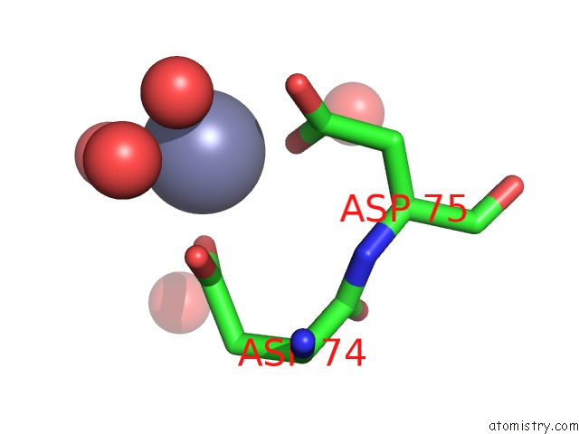
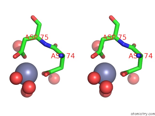
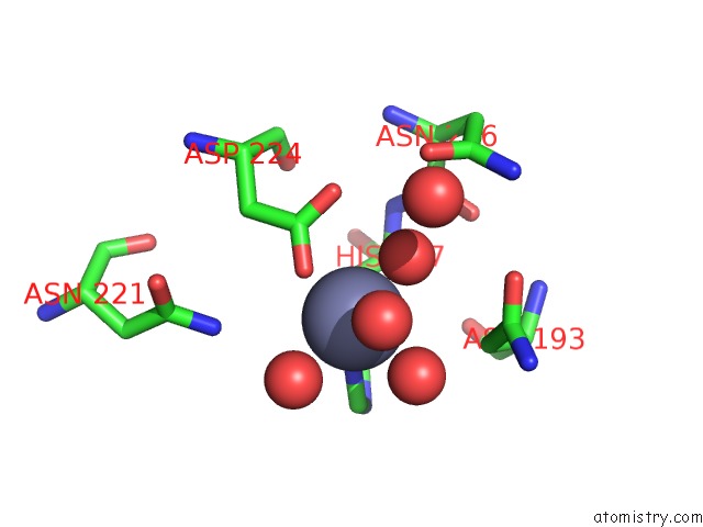
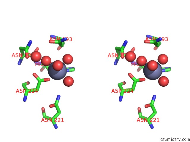
Zinc binding site 2 out of 6 in 3ea2
Go back to
Zinc binding site 2 out
of 6 in the Crystal Structure of the Myo-Inositol Bound Y247S/Y251S Mutant of Phosphatidylinositol-Specific Phospholipase C From Bacillus Thuringiensis
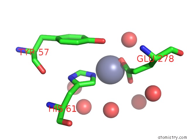
Mono view
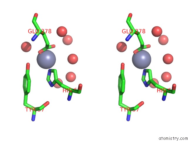
Stereo pair view
Mono view
Stereo pair view
A full contact list of Zinc with other atoms in the Zn binding
site number 2 of Crystal Structure of the Myo-Inositol Bound Y247S/Y251S Mutant of Phosphatidylinositol-Specific Phospholipase C From Bacillus Thuringiensis within 5.0Å range:
|
Zinc binding site 3 out of 6 in 3ea2
Go back to
Zinc binding site 3 out
of 6 in the Crystal Structure of the Myo-Inositol Bound Y247S/Y251S Mutant of Phosphatidylinositol-Specific Phospholipase C From Bacillus Thuringiensis
Mono view
Stereo pair view
Mono view
Stereo pair view
A full contact list of Zinc with other atoms in the Zn binding
site number 3 of Crystal Structure of the Myo-Inositol Bound Y247S/Y251S Mutant of Phosphatidylinositol-Specific Phospholipase C From Bacillus Thuringiensis within 5.0Å range:
|
Zinc binding site 4 out of 6 in 3ea2
Go back to
Zinc binding site 4 out
of 6 in the Crystal Structure of the Myo-Inositol Bound Y247S/Y251S Mutant of Phosphatidylinositol-Specific Phospholipase C From Bacillus Thuringiensis
Mono view
Stereo pair view
Mono view
Stereo pair view
A full contact list of Zinc with other atoms in the Zn binding
site number 4 of Crystal Structure of the Myo-Inositol Bound Y247S/Y251S Mutant of Phosphatidylinositol-Specific Phospholipase C From Bacillus Thuringiensis within 5.0Å range:
|
Zinc binding site 5 out of 6 in 3ea2
Go back to
Zinc binding site 5 out
of 6 in the Crystal Structure of the Myo-Inositol Bound Y247S/Y251S Mutant of Phosphatidylinositol-Specific Phospholipase C From Bacillus Thuringiensis
Mono view
Stereo pair view
Mono view
Stereo pair view
A full contact list of Zinc with other atoms in the Zn binding
site number 5 of Crystal Structure of the Myo-Inositol Bound Y247S/Y251S Mutant of Phosphatidylinositol-Specific Phospholipase C From Bacillus Thuringiensis within 5.0Å range:
|
Zinc binding site 6 out of 6 in 3ea2
Go back to
Zinc binding site 6 out
of 6 in the Crystal Structure of the Myo-Inositol Bound Y247S/Y251S Mutant of Phosphatidylinositol-Specific Phospholipase C From Bacillus Thuringiensis
Mono view
Stereo pair view
Mono view
Stereo pair view
A full contact list of Zinc with other atoms in the Zn binding
site number 6 of Crystal Structure of the Myo-Inositol Bound Y247S/Y251S Mutant of Phosphatidylinositol-Specific Phospholipase C From Bacillus Thuringiensis within 5.0Å range:
|
Reference:
X.Shi,
C.Shao,
X.Zhang,
C.Zambonelli,
A.G.Redfield,
J.F.Head,
B.A.Seaton,
M.F.Roberts.
Modulation of Bacillus Thuringiensis Phosphatidylinositol-Specific Phospholipase C Activity By Mutations in the Putative Dimerization Interface. J.Biol.Chem. V. 284 15607 2009.
ISSN: ISSN 0021-9258
PubMed: 19369255
DOI: 10.1074/JBC.M901601200
Page generated: Thu Oct 24 12:41:29 2024
ISSN: ISSN 0021-9258
PubMed: 19369255
DOI: 10.1074/JBC.M901601200
Last articles
Zn in 9J0NZn in 9J0O
Zn in 9J0P
Zn in 9FJX
Zn in 9EKB
Zn in 9C0F
Zn in 9CAH
Zn in 9CH0
Zn in 9CH3
Zn in 9CH1