Zinc »
PDB 3e2u-3eb9 »
3e80 »
Zinc in PDB 3e80: Structure of Heparinase II Complexed with Heparan Sulfate Degradation Disaccharide Product
Protein crystallography data
The structure of Structure of Heparinase II Complexed with Heparan Sulfate Degradation Disaccharide Product, PDB code: 3e80
was solved by
D.Shaya,
M.Cygler,
with X-Ray Crystallography technique. A brief refinement statistics is given in the table below:
| Resolution Low / High (Å) | 37.57 / 2.35 |
| Space group | P 21 21 2 |
| Cell size a, b, c (Å), α, β, γ (°) | 201.280, 209.360, 59.220, 90.00, 90.00, 90.00 |
| R / Rfree (%) | 22.9 / 26.7 |
Zinc Binding Sites:
The binding sites of Zinc atom in the Structure of Heparinase II Complexed with Heparan Sulfate Degradation Disaccharide Product
(pdb code 3e80). This binding sites where shown within
5.0 Angstroms radius around Zinc atom.
In total 3 binding sites of Zinc where determined in the Structure of Heparinase II Complexed with Heparan Sulfate Degradation Disaccharide Product, PDB code: 3e80:
Jump to Zinc binding site number: 1; 2; 3;
In total 3 binding sites of Zinc where determined in the Structure of Heparinase II Complexed with Heparan Sulfate Degradation Disaccharide Product, PDB code: 3e80:
Jump to Zinc binding site number: 1; 2; 3;
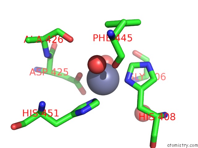
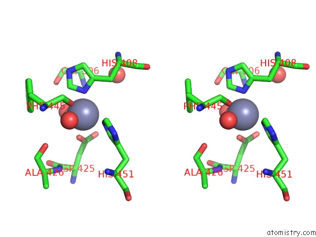
Zinc binding site 1 out of 3 in 3e80
Go back to
Zinc binding site 1 out
of 3 in the Structure of Heparinase II Complexed with Heparan Sulfate Degradation Disaccharide Product
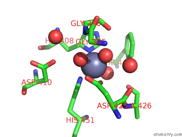
Mono view
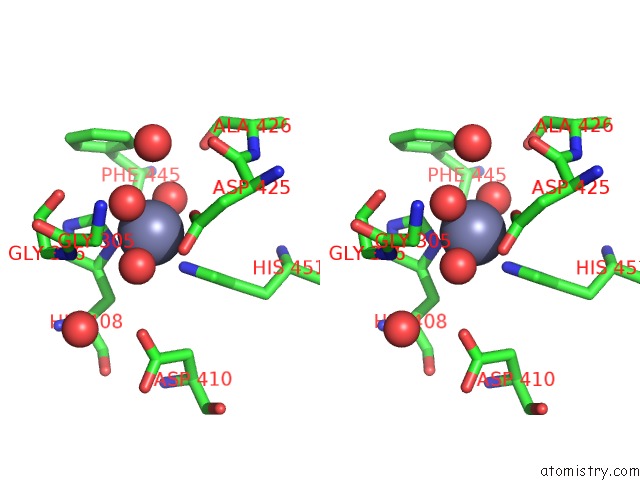
Stereo pair view
Mono view
Stereo pair view
A full contact list of Zinc with other atoms in the Zn binding
site number 1 of Structure of Heparinase II Complexed with Heparan Sulfate Degradation Disaccharide Product within 5.0Å range:
|
Zinc binding site 2 out of 3 in 3e80
Go back to
Zinc binding site 2 out
of 3 in the Structure of Heparinase II Complexed with Heparan Sulfate Degradation Disaccharide Product
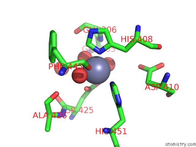
Mono view
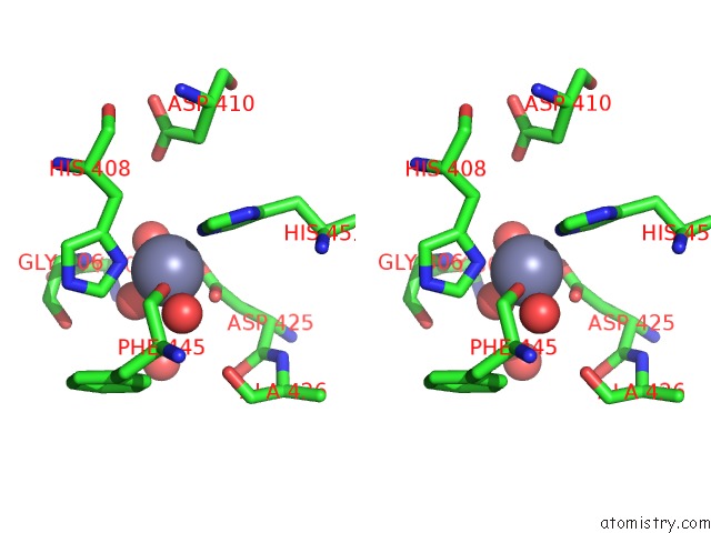
Stereo pair view
Mono view
Stereo pair view
A full contact list of Zinc with other atoms in the Zn binding
site number 2 of Structure of Heparinase II Complexed with Heparan Sulfate Degradation Disaccharide Product within 5.0Å range:
|
Zinc binding site 3 out of 3 in 3e80
Go back to
Zinc binding site 3 out
of 3 in the Structure of Heparinase II Complexed with Heparan Sulfate Degradation Disaccharide Product
Mono view
Stereo pair view
Mono view
Stereo pair view
A full contact list of Zinc with other atoms in the Zn binding
site number 3 of Structure of Heparinase II Complexed with Heparan Sulfate Degradation Disaccharide Product within 5.0Å range:
|
Reference:
D.Shaya,
W.Zhao,
M.L.Garron,
Z.Xiao,
Q.Cui,
Z.Zhang,
T.Sulea,
R.J.Linhardt,
M.Cygler.
Catalytic Mechanism of Heparinase II Investigated By Site-Directed Mutagenesis and the Crystal Structure with Its Substrate. J.Biol.Chem. V. 285 20051 2010.
ISSN: ISSN 0021-9258
PubMed: 20404324
DOI: 10.1074/JBC.M110.101071
Page generated: Thu Oct 24 12:40:47 2024
ISSN: ISSN 0021-9258
PubMed: 20404324
DOI: 10.1074/JBC.M110.101071
Last articles
Zn in 9J0NZn in 9J0O
Zn in 9J0P
Zn in 9FJX
Zn in 9EKB
Zn in 9C0F
Zn in 9CAH
Zn in 9CH0
Zn in 9CH3
Zn in 9CH1