Zinc »
PDB 3e2u-3eb9 »
3e73 »
Zinc in PDB 3e73: Crystal Structure of Human LANCL1 Complexed with Gsh
Protein crystallography data
The structure of Crystal Structure of Human LANCL1 Complexed with Gsh, PDB code: 3e73
was solved by
W.Zhang,
G.Zhu,
X.Li,
Z.Rao,
C.Zhang,
with X-Ray Crystallography technique. A brief refinement statistics is given in the table below:
| Resolution Low / High (Å) | 49.33 / 2.80 |
| Space group | P 65 2 2 |
| Cell size a, b, c (Å), α, β, γ (°) | 194.100, 194.101, 243.554, 90.00, 90.00, 120.00 |
| R / Rfree (%) | 23.1 / 26.2 |
Zinc Binding Sites:
The binding sites of Zinc atom in the Crystal Structure of Human LANCL1 Complexed with Gsh
(pdb code 3e73). This binding sites where shown within
5.0 Angstroms radius around Zinc atom.
In total 2 binding sites of Zinc where determined in the Crystal Structure of Human LANCL1 Complexed with Gsh, PDB code: 3e73:
Jump to Zinc binding site number: 1; 2;
In total 2 binding sites of Zinc where determined in the Crystal Structure of Human LANCL1 Complexed with Gsh, PDB code: 3e73:
Jump to Zinc binding site number: 1; 2;
Zinc binding site 1 out of 2 in 3e73
Go back to
Zinc binding site 1 out
of 2 in the Crystal Structure of Human LANCL1 Complexed with Gsh
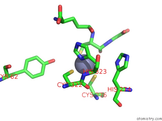
Mono view
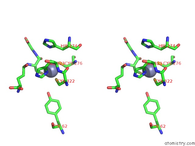
Stereo pair view
Mono view
Stereo pair view
A full contact list of Zinc with other atoms in the Zn binding
site number 1 of Crystal Structure of Human LANCL1 Complexed with Gsh within 5.0Å range:
|
Zinc binding site 2 out of 2 in 3e73
Go back to
Zinc binding site 2 out
of 2 in the Crystal Structure of Human LANCL1 Complexed with Gsh
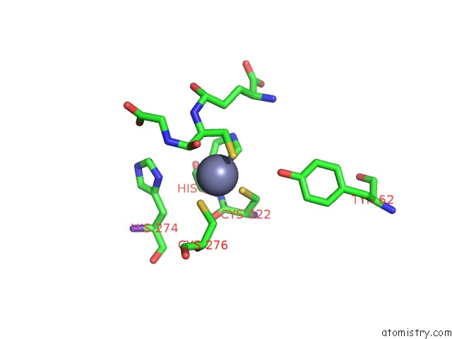
Mono view
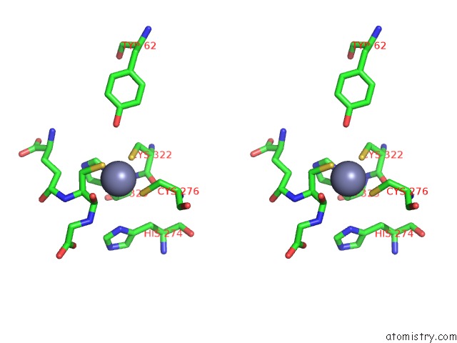
Stereo pair view
Mono view
Stereo pair view
A full contact list of Zinc with other atoms in the Zn binding
site number 2 of Crystal Structure of Human LANCL1 Complexed with Gsh within 5.0Å range:
|
Reference:
W.Zhang,
L.Wang,
Y.Liu,
J.Xu,
G.Zhu,
H.Cang,
X.Li,
M.Bartlam,
K.Hensley,
G.Li,
Z.Rao,
X.C.Zhang.
Structure of Human Lanthionine Synthetase C-Like Protein 1 and Its Interaction with EPS8 and Glutathione Genes Dev. V. 23 1387 2009.
ISSN: ISSN 0890-9369
PubMed: 19528316
DOI: 10.1101/GAD.1789209
Page generated: Thu Oct 24 12:38:52 2024
ISSN: ISSN 0890-9369
PubMed: 19528316
DOI: 10.1101/GAD.1789209
Last articles
Zn in 9J0NZn in 9J0O
Zn in 9J0P
Zn in 9FJX
Zn in 9EKB
Zn in 9C0F
Zn in 9CAH
Zn in 9CH0
Zn in 9CH3
Zn in 9CH1