Zinc »
PDB 3e2u-3eb9 »
3e4z »
Zinc in PDB 3e4z: Crystal Structure of Human Insulin Degrading Enzyme in Complex with Insulin-Like Growth Factor II
Enzymatic activity of Crystal Structure of Human Insulin Degrading Enzyme in Complex with Insulin-Like Growth Factor II
All present enzymatic activity of Crystal Structure of Human Insulin Degrading Enzyme in Complex with Insulin-Like Growth Factor II:
3.4.24.56;
3.4.24.56;
Protein crystallography data
The structure of Crystal Structure of Human Insulin Degrading Enzyme in Complex with Insulin-Like Growth Factor II, PDB code: 3e4z
was solved by
Q.Guo,
M.Manolopoulou,
W.-J.Tang,
with X-Ray Crystallography technique. A brief refinement statistics is given in the table below:
| Resolution Low / High (Å) | 50.00 / 2.28 |
| Space group | P 65 |
| Cell size a, b, c (Å), α, β, γ (°) | 263.033, 263.033, 90.816, 90.00, 90.00, 120.00 |
| R / Rfree (%) | 19.6 / 23 |
Zinc Binding Sites:
The binding sites of Zinc atom in the Crystal Structure of Human Insulin Degrading Enzyme in Complex with Insulin-Like Growth Factor II
(pdb code 3e4z). This binding sites where shown within
5.0 Angstroms radius around Zinc atom.
In total 2 binding sites of Zinc where determined in the Crystal Structure of Human Insulin Degrading Enzyme in Complex with Insulin-Like Growth Factor II, PDB code: 3e4z:
Jump to Zinc binding site number: 1; 2;
In total 2 binding sites of Zinc where determined in the Crystal Structure of Human Insulin Degrading Enzyme in Complex with Insulin-Like Growth Factor II, PDB code: 3e4z:
Jump to Zinc binding site number: 1; 2;
Zinc binding site 1 out of 2 in 3e4z
Go back to
Zinc binding site 1 out
of 2 in the Crystal Structure of Human Insulin Degrading Enzyme in Complex with Insulin-Like Growth Factor II
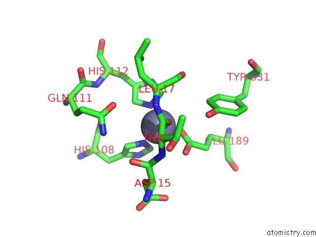
Mono view
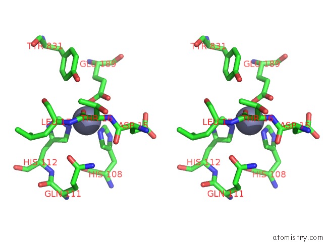
Stereo pair view
Mono view
Stereo pair view
A full contact list of Zinc with other atoms in the Zn binding
site number 1 of Crystal Structure of Human Insulin Degrading Enzyme in Complex with Insulin-Like Growth Factor II within 5.0Å range:
|
Zinc binding site 2 out of 2 in 3e4z
Go back to
Zinc binding site 2 out
of 2 in the Crystal Structure of Human Insulin Degrading Enzyme in Complex with Insulin-Like Growth Factor II
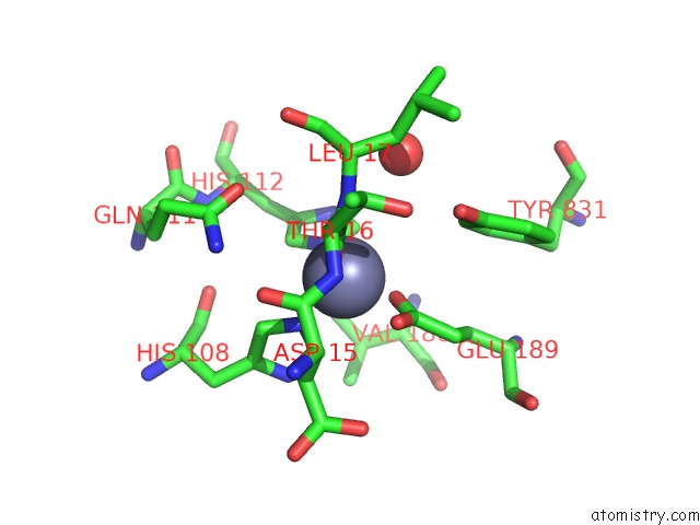
Mono view
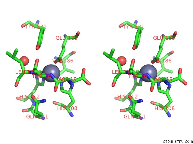
Stereo pair view
Mono view
Stereo pair view
A full contact list of Zinc with other atoms in the Zn binding
site number 2 of Crystal Structure of Human Insulin Degrading Enzyme in Complex with Insulin-Like Growth Factor II within 5.0Å range:
|
Reference:
Q.Guo,
M.Manolopoulou,
Y.Bian,
A.B.Schilling,
W.J.Tang.
Molecular Basis For the Recognition and Cleavages of Igf-II, Tgf-Alpha, and Amylin By Human Insulin-Degrading Enzyme. J.Mol.Biol. V. 395 430 2010.
ISSN: ISSN 0022-2836
PubMed: 19896952
DOI: 10.1016/J.JMB.2009.10.072
Page generated: Thu Oct 24 12:38:07 2024
ISSN: ISSN 0022-2836
PubMed: 19896952
DOI: 10.1016/J.JMB.2009.10.072
Last articles
Zn in 9J0NZn in 9J0O
Zn in 9J0P
Zn in 9FJX
Zn in 9EKB
Zn in 9C0F
Zn in 9CAH
Zn in 9CH0
Zn in 9CH3
Zn in 9CH1