Zinc »
PDB 3e2u-3eb9 »
3e2w »
Zinc in PDB 3e2w: H. Influenzae Beta-Carbonic Anhydrase, Variant Y181F with 1M Bicarbonate
Enzymatic activity of H. Influenzae Beta-Carbonic Anhydrase, Variant Y181F with 1M Bicarbonate
All present enzymatic activity of H. Influenzae Beta-Carbonic Anhydrase, Variant Y181F with 1M Bicarbonate:
4.2.1.1;
4.2.1.1;
Protein crystallography data
The structure of H. Influenzae Beta-Carbonic Anhydrase, Variant Y181F with 1M Bicarbonate, PDB code: 3e2w
was solved by
R.S.Rowlett,
J.Lee,
with X-Ray Crystallography technique. A brief refinement statistics is given in the table below:
| Resolution Low / High (Å) | 26.20 / 2.30 |
| Space group | C 1 2 1 |
| Cell size a, b, c (Å), α, β, γ (°) | 250.141, 145.225, 53.499, 90.00, 93.78, 90.00 |
| R / Rfree (%) | 20.5 / 24.2 |
Zinc Binding Sites:
The binding sites of Zinc atom in the H. Influenzae Beta-Carbonic Anhydrase, Variant Y181F with 1M Bicarbonate
(pdb code 3e2w). This binding sites where shown within
5.0 Angstroms radius around Zinc atom.
In total 6 binding sites of Zinc where determined in the H. Influenzae Beta-Carbonic Anhydrase, Variant Y181F with 1M Bicarbonate, PDB code: 3e2w:
Jump to Zinc binding site number: 1; 2; 3; 4; 5; 6;
In total 6 binding sites of Zinc where determined in the H. Influenzae Beta-Carbonic Anhydrase, Variant Y181F with 1M Bicarbonate, PDB code: 3e2w:
Jump to Zinc binding site number: 1; 2; 3; 4; 5; 6;
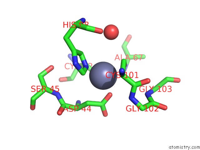
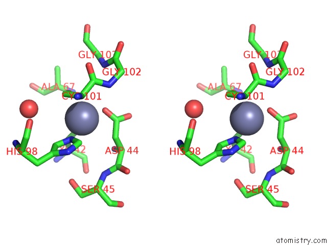
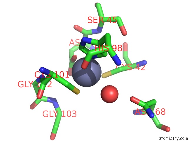
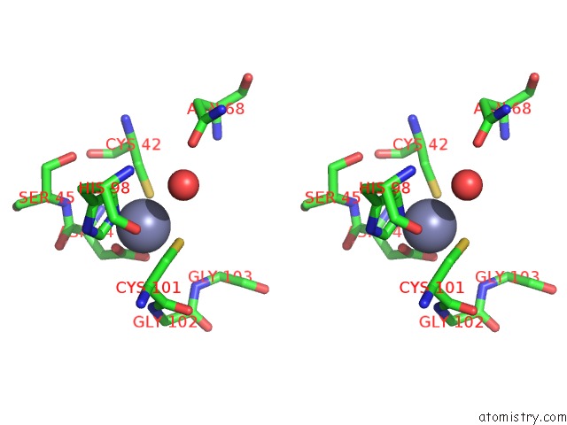
Zinc binding site 1 out of 6 in 3e2w
Go back to
Zinc binding site 1 out
of 6 in the H. Influenzae Beta-Carbonic Anhydrase, Variant Y181F with 1M Bicarbonate
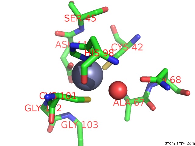
Mono view
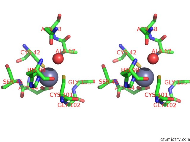
Stereo pair view
Mono view
Stereo pair view
A full contact list of Zinc with other atoms in the Zn binding
site number 1 of H. Influenzae Beta-Carbonic Anhydrase, Variant Y181F with 1M Bicarbonate within 5.0Å range:
|
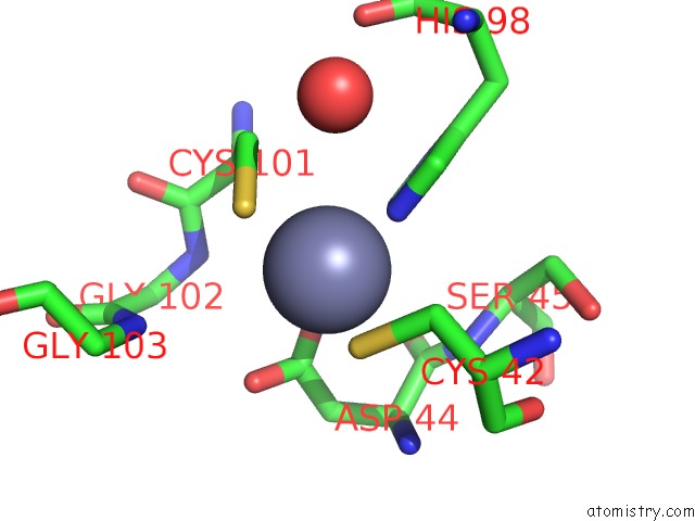
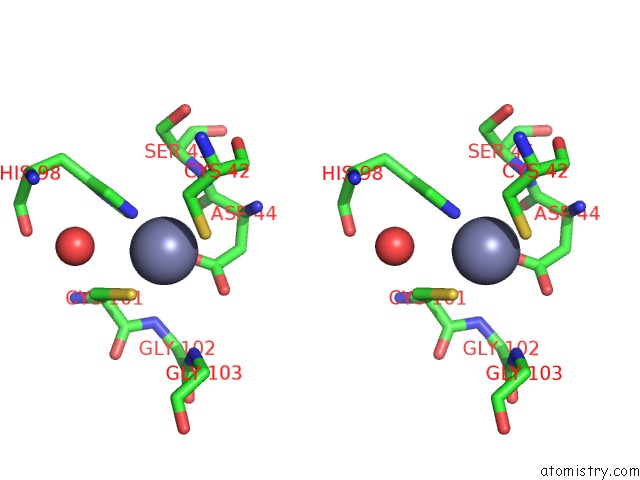
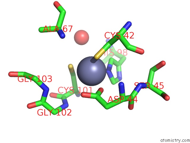
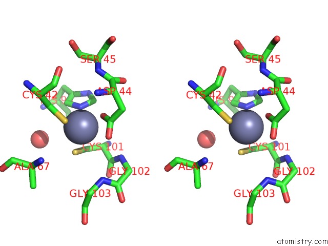
Zinc binding site 2 out of 6 in 3e2w
Go back to
Zinc binding site 2 out
of 6 in the H. Influenzae Beta-Carbonic Anhydrase, Variant Y181F with 1M Bicarbonate
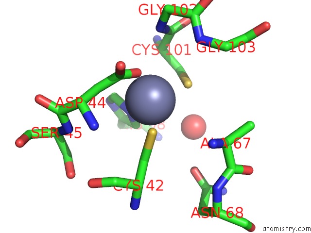
Mono view
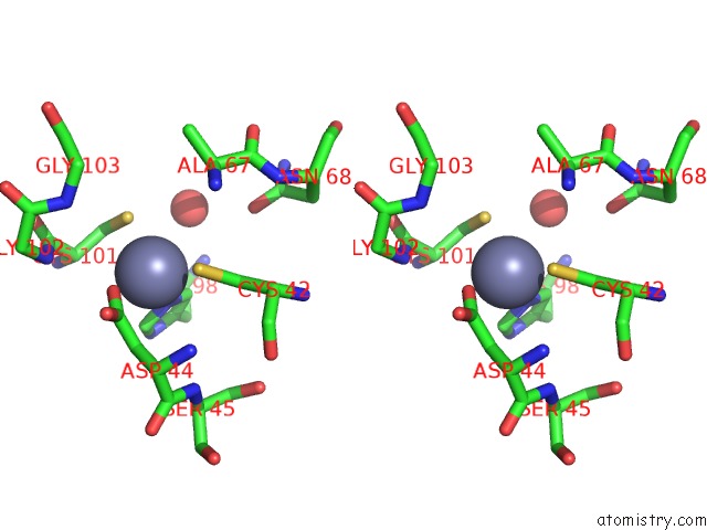
Stereo pair view
Mono view
Stereo pair view
A full contact list of Zinc with other atoms in the Zn binding
site number 2 of H. Influenzae Beta-Carbonic Anhydrase, Variant Y181F with 1M Bicarbonate within 5.0Å range:
|
Zinc binding site 3 out of 6 in 3e2w
Go back to
Zinc binding site 3 out
of 6 in the H. Influenzae Beta-Carbonic Anhydrase, Variant Y181F with 1M Bicarbonate
Mono view
Stereo pair view
Mono view
Stereo pair view
A full contact list of Zinc with other atoms in the Zn binding
site number 3 of H. Influenzae Beta-Carbonic Anhydrase, Variant Y181F with 1M Bicarbonate within 5.0Å range:
|
Zinc binding site 4 out of 6 in 3e2w
Go back to
Zinc binding site 4 out
of 6 in the H. Influenzae Beta-Carbonic Anhydrase, Variant Y181F with 1M Bicarbonate
Mono view
Stereo pair view
Mono view
Stereo pair view
A full contact list of Zinc with other atoms in the Zn binding
site number 4 of H. Influenzae Beta-Carbonic Anhydrase, Variant Y181F with 1M Bicarbonate within 5.0Å range:
|
Zinc binding site 5 out of 6 in 3e2w
Go back to
Zinc binding site 5 out
of 6 in the H. Influenzae Beta-Carbonic Anhydrase, Variant Y181F with 1M Bicarbonate
Mono view
Stereo pair view
Mono view
Stereo pair view
A full contact list of Zinc with other atoms in the Zn binding
site number 5 of H. Influenzae Beta-Carbonic Anhydrase, Variant Y181F with 1M Bicarbonate within 5.0Å range:
|
Zinc binding site 6 out of 6 in 3e2w
Go back to
Zinc binding site 6 out
of 6 in the H. Influenzae Beta-Carbonic Anhydrase, Variant Y181F with 1M Bicarbonate
Mono view
Stereo pair view
Mono view
Stereo pair view
A full contact list of Zinc with other atoms in the Zn binding
site number 6 of H. Influenzae Beta-Carbonic Anhydrase, Variant Y181F with 1M Bicarbonate within 5.0Å range:
|
Reference:
R.S.Rowlett,
C.Tu,
J.Lee,
A.G.Herman,
D.A.Chapnick,
S.H.Shah,
P.C.Gareiss.
Allosteric Site Variants of Haemophilus Influenzae Beta-Carbonic Anhydrase. Biochemistry V. 48 6146 2009.
ISSN: ISSN 0006-2960
PubMed: 19459702
DOI: 10.1021/BI900663H
Page generated: Thu Oct 24 12:33:39 2024
ISSN: ISSN 0006-2960
PubMed: 19459702
DOI: 10.1021/BI900663H
Last articles
Zn in 9J0NZn in 9J0O
Zn in 9J0P
Zn in 9FJX
Zn in 9EKB
Zn in 9C0F
Zn in 9CAH
Zn in 9CH0
Zn in 9CH3
Zn in 9CH1