Zinc »
PDB 3dgn-3dsw »
3dow »
Zinc in PDB 3dow: Complex Structure of Gaba Type A Receptor Associated Protein and Its Binding Epitope on Calreticulin
Protein crystallography data
The structure of Complex Structure of Gaba Type A Receptor Associated Protein and Its Binding Epitope on Calreticulin, PDB code: 3dow
was solved by
Y.Thielmann,
O.H.Weiergraeber,
D.Willbold,
with X-Ray Crystallography technique. A brief refinement statistics is given in the table below:
| Resolution Low / High (Å) | 34.31 / 2.30 |
| Space group | I 2 3 |
| Cell size a, b, c (Å), α, β, γ (°) | 97.043, 97.043, 97.043, 90.00, 90.00, 90.00 |
| R / Rfree (%) | 23.2 / 27 |
Zinc Binding Sites:
The binding sites of Zinc atom in the Complex Structure of Gaba Type A Receptor Associated Protein and Its Binding Epitope on Calreticulin
(pdb code 3dow). This binding sites where shown within
5.0 Angstroms radius around Zinc atom.
In total only one binding site of Zinc was determined in the Complex Structure of Gaba Type A Receptor Associated Protein and Its Binding Epitope on Calreticulin, PDB code: 3dow:
In total only one binding site of Zinc was determined in the Complex Structure of Gaba Type A Receptor Associated Protein and Its Binding Epitope on Calreticulin, PDB code: 3dow:
Zinc binding site 1 out of 1 in 3dow
Go back to
Zinc binding site 1 out
of 1 in the Complex Structure of Gaba Type A Receptor Associated Protein and Its Binding Epitope on Calreticulin
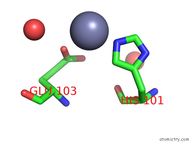
Mono view
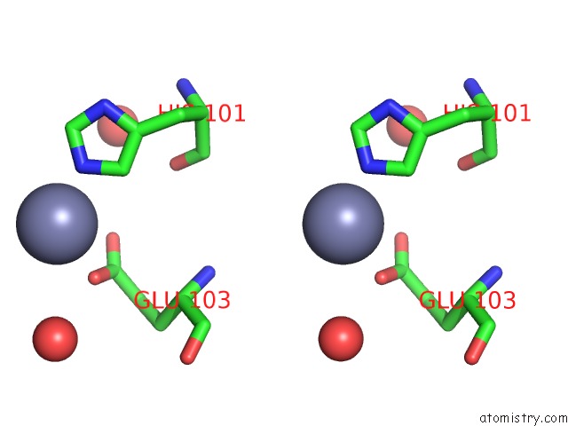
Stereo pair view
Mono view
Stereo pair view
A full contact list of Zinc with other atoms in the Zn binding
site number 1 of Complex Structure of Gaba Type A Receptor Associated Protein and Its Binding Epitope on Calreticulin within 5.0Å range:
|
Reference:
Y.Thielmann,
O.H.Weiergraber,
J.Mohrluder,
D.Willbold.
Structural Framework of the Gabarap-Calreticulin Interface - Implications For Substrate Binding to Endoplasmic Reticulum Chaperones. Febs J. V. 276 1140 2009.
ISSN: ISSN 1742-464X
PubMed: 19154346
DOI: 10.1111/J.1742-4658.2008.06857.X
Page generated: Thu Oct 24 12:17:00 2024
ISSN: ISSN 1742-464X
PubMed: 19154346
DOI: 10.1111/J.1742-4658.2008.06857.X
Last articles
Zn in 9MJ5Zn in 9HNW
Zn in 9G0L
Zn in 9FNE
Zn in 9DZN
Zn in 9E0I
Zn in 9D32
Zn in 9DAK
Zn in 8ZXC
Zn in 8ZUF