Zinc »
PDB 3cjp-3czs »
3cww »
Zinc in PDB 3cww: Crystal Structure of Ide-Bradykinin Complex
Enzymatic activity of Crystal Structure of Ide-Bradykinin Complex
All present enzymatic activity of Crystal Structure of Ide-Bradykinin Complex:
3.4.24.56;
3.4.24.56;
Protein crystallography data
The structure of Crystal Structure of Ide-Bradykinin Complex, PDB code: 3cww
was solved by
E.Malito,
W.J.Tang,
with X-Ray Crystallography technique. A brief refinement statistics is given in the table below:
| Resolution Low / High (Å) | 29.77 / 1.96 |
| Space group | P 65 |
| Cell size a, b, c (Å), α, β, γ (°) | 262.448, 262.448, 90.628, 90.00, 90.00, 120.00 |
| R / Rfree (%) | 18.1 / 20.8 |
Zinc Binding Sites:
The binding sites of Zinc atom in the Crystal Structure of Ide-Bradykinin Complex
(pdb code 3cww). This binding sites where shown within
5.0 Angstroms radius around Zinc atom.
In total 2 binding sites of Zinc where determined in the Crystal Structure of Ide-Bradykinin Complex, PDB code: 3cww:
Jump to Zinc binding site number: 1; 2;
In total 2 binding sites of Zinc where determined in the Crystal Structure of Ide-Bradykinin Complex, PDB code: 3cww:
Jump to Zinc binding site number: 1; 2;
Zinc binding site 1 out of 2 in 3cww
Go back to
Zinc binding site 1 out
of 2 in the Crystal Structure of Ide-Bradykinin Complex
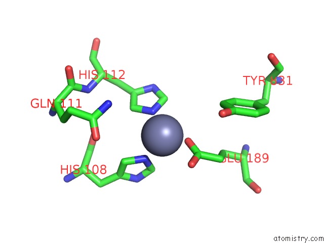
Mono view
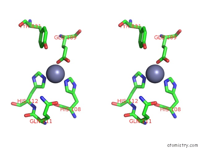
Stereo pair view
Mono view
Stereo pair view
A full contact list of Zinc with other atoms in the Zn binding
site number 1 of Crystal Structure of Ide-Bradykinin Complex within 5.0Å range:
|
Zinc binding site 2 out of 2 in 3cww
Go back to
Zinc binding site 2 out
of 2 in the Crystal Structure of Ide-Bradykinin Complex
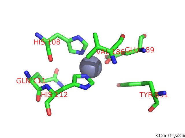
Mono view
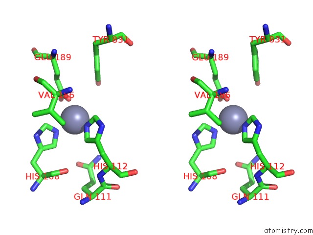
Stereo pair view
Mono view
Stereo pair view
A full contact list of Zinc with other atoms in the Zn binding
site number 2 of Crystal Structure of Ide-Bradykinin Complex within 5.0Å range:
|
Reference:
E.Malito,
L.A.Ralat,
M.Manolopoulou,
J.L.Tsay,
N.L.Wadlington,
W.J.Tang.
Molecular Bases For the Recognition of Short Peptide Substrates and Cysteine-Directed Modifications of Human Insulin-Degrading Enzyme Biochemistry V. 47 12822 2008.
ISSN: ISSN 0006-2960
PubMed: 18986166
DOI: 10.1021/BI801192H
Page generated: Thu Oct 24 11:57:51 2024
ISSN: ISSN 0006-2960
PubMed: 18986166
DOI: 10.1021/BI801192H
Last articles
Zn in 9J0NZn in 9J0O
Zn in 9J0P
Zn in 9FJX
Zn in 9EKB
Zn in 9C0F
Zn in 9CAH
Zn in 9CH0
Zn in 9CH3
Zn in 9CH1