Zinc »
PDB 3cjp-3czs »
3cmr »
Zinc in PDB 3cmr: E. Coli Alkaline Phosphatase Mutant R166S in Complex with Phosphate
Enzymatic activity of E. Coli Alkaline Phosphatase Mutant R166S in Complex with Phosphate
All present enzymatic activity of E. Coli Alkaline Phosphatase Mutant R166S in Complex with Phosphate:
3.1.3.1;
3.1.3.1;
Protein crystallography data
The structure of E. Coli Alkaline Phosphatase Mutant R166S in Complex with Phosphate, PDB code: 3cmr
was solved by
P.J.O'brien,
J.K.Lassila,
T.D.Fenn,
J.G.Zalatan,
D.Herschlag,
with X-Ray Crystallography technique. A brief refinement statistics is given in the table below:
| Resolution Low / High (Å) | 38.75 / 2.05 |
| Space group | P 63 2 2 |
| Cell size a, b, c (Å), α, β, γ (°) | 160.614, 160.614, 139.923, 90.00, 90.00, 120.00 |
| R / Rfree (%) | 18 / 21.3 |
Other elements in 3cmr:
The structure of E. Coli Alkaline Phosphatase Mutant R166S in Complex with Phosphate also contains other interesting chemical elements:
| Magnesium | (Mg) | 2 atoms |
Zinc Binding Sites:
The binding sites of Zinc atom in the E. Coli Alkaline Phosphatase Mutant R166S in Complex with Phosphate
(pdb code 3cmr). This binding sites where shown within
5.0 Angstroms radius around Zinc atom.
In total 4 binding sites of Zinc where determined in the E. Coli Alkaline Phosphatase Mutant R166S in Complex with Phosphate, PDB code: 3cmr:
Jump to Zinc binding site number: 1; 2; 3; 4;
In total 4 binding sites of Zinc where determined in the E. Coli Alkaline Phosphatase Mutant R166S in Complex with Phosphate, PDB code: 3cmr:
Jump to Zinc binding site number: 1; 2; 3; 4;
Zinc binding site 1 out of 4 in 3cmr
Go back to
Zinc binding site 1 out
of 4 in the E. Coli Alkaline Phosphatase Mutant R166S in Complex with Phosphate

Mono view
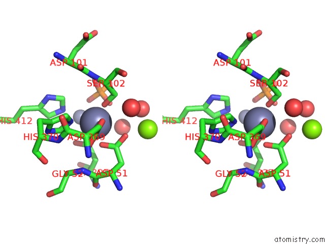
Stereo pair view
Mono view
Stereo pair view
A full contact list of Zinc with other atoms in the Zn binding
site number 1 of E. Coli Alkaline Phosphatase Mutant R166S in Complex with Phosphate within 5.0Å range:
|
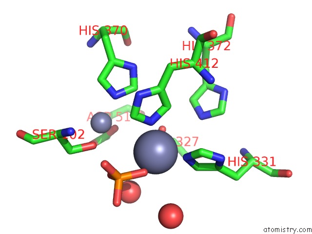
Zinc binding site 2 out of 4 in 3cmr
Go back to
Zinc binding site 2 out
of 4 in the E. Coli Alkaline Phosphatase Mutant R166S in Complex with Phosphate
Mono view

Stereo pair view
Mono view
Stereo pair view
A full contact list of Zinc with other atoms in the Zn binding
site number 2 of E. Coli Alkaline Phosphatase Mutant R166S in Complex with Phosphate within 5.0Å range:
|
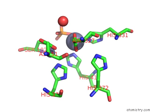
Zinc binding site 3 out of 4 in 3cmr
Go back to
Zinc binding site 3 out
of 4 in the E. Coli Alkaline Phosphatase Mutant R166S in Complex with Phosphate

Mono view

Stereo pair view
Mono view
Stereo pair view
A full contact list of Zinc with other atoms in the Zn binding
site number 3 of E. Coli Alkaline Phosphatase Mutant R166S in Complex with Phosphate within 5.0Å range:
|
Zinc binding site 4 out of 4 in 3cmr
Go back to
Zinc binding site 4 out
of 4 in the E. Coli Alkaline Phosphatase Mutant R166S in Complex with Phosphate
Mono view
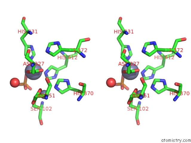
Stereo pair view
Mono view
Stereo pair view
A full contact list of Zinc with other atoms in the Zn binding
site number 4 of E. Coli Alkaline Phosphatase Mutant R166S in Complex with Phosphate within 5.0Å range:
|
Reference:
P.J.O'brien,
J.K.Lassila,
T.D.Fenn,
J.G.Zalatan,
D.Herschlag.
Arginine Coordination in Enzymatic Phosphoryl Transfer: Evaluation of the Effect of ARG166 Mutations in Escherichia Coli Alkaline Phosphatase Biochemistry V. 47 7663 2008.
ISSN: ISSN 0006-2960
PubMed: 18627128
DOI: 10.1021/BI800545N
Page generated: Thu Oct 24 11:51:29 2024
ISSN: ISSN 0006-2960
PubMed: 18627128
DOI: 10.1021/BI800545N
Last articles
Zn in 9J0NZn in 9J0O
Zn in 9J0P
Zn in 9FJX
Zn in 9EKB
Zn in 9C0F
Zn in 9CAH
Zn in 9CH0
Zn in 9CH3
Zn in 9CH1