Zinc »
PDB 3c52-3cjj »
3c58 »
Zinc in PDB 3c58: Crystal Structure of A Complex Between the Wild-Type Lactococcus Lactis Fpg (Mutm) and A N7-Benzyl-Fapy-Dg Containing Dna
Enzymatic activity of Crystal Structure of A Complex Between the Wild-Type Lactococcus Lactis Fpg (Mutm) and A N7-Benzyl-Fapy-Dg Containing Dna
All present enzymatic activity of Crystal Structure of A Complex Between the Wild-Type Lactococcus Lactis Fpg (Mutm) and A N7-Benzyl-Fapy-Dg Containing Dna:
4.2.99.18;
4.2.99.18;
Protein crystallography data
The structure of Crystal Structure of A Complex Between the Wild-Type Lactococcus Lactis Fpg (Mutm) and A N7-Benzyl-Fapy-Dg Containing Dna, PDB code: 3c58
was solved by
F.Coste,
B.Castaing,
T.Carell,
with X-Ray Crystallography technique. A brief refinement statistics is given in the table below:
| Resolution Low / High (Å) | 45.69 / 1.90 |
| Space group | P 41 21 2 |
| Cell size a, b, c (Å), α, β, γ (°) | 91.356, 91.356, 140.943, 90.00, 90.00, 90.00 |
| R / Rfree (%) | 17.2 / 20 |
Zinc Binding Sites:
The binding sites of Zinc atom in the Crystal Structure of A Complex Between the Wild-Type Lactococcus Lactis Fpg (Mutm) and A N7-Benzyl-Fapy-Dg Containing Dna
(pdb code 3c58). This binding sites where shown within
5.0 Angstroms radius around Zinc atom.
In total only one binding site of Zinc was determined in the Crystal Structure of A Complex Between the Wild-Type Lactococcus Lactis Fpg (Mutm) and A N7-Benzyl-Fapy-Dg Containing Dna, PDB code: 3c58:
In total only one binding site of Zinc was determined in the Crystal Structure of A Complex Between the Wild-Type Lactococcus Lactis Fpg (Mutm) and A N7-Benzyl-Fapy-Dg Containing Dna, PDB code: 3c58:
Zinc binding site 1 out of 1 in 3c58
Go back to
Zinc binding site 1 out
of 1 in the Crystal Structure of A Complex Between the Wild-Type Lactococcus Lactis Fpg (Mutm) and A N7-Benzyl-Fapy-Dg Containing Dna
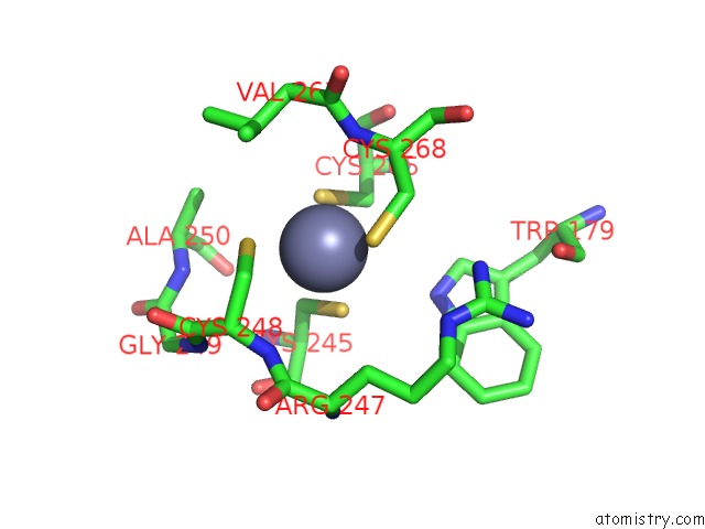
Mono view
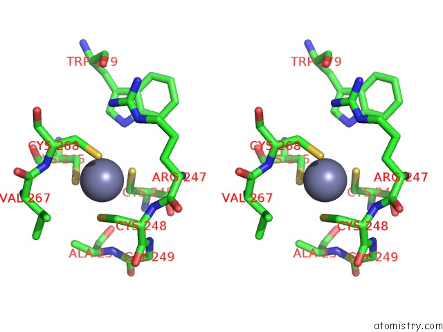
Stereo pair view
Mono view
Stereo pair view
A full contact list of Zinc with other atoms in the Zn binding
site number 1 of Crystal Structure of A Complex Between the Wild-Type Lactococcus Lactis Fpg (Mutm) and A N7-Benzyl-Fapy-Dg Containing Dna within 5.0Å range:
|
Reference:
F.Coste,
M.Ober,
Y.V.Le Bihan,
M.A.Izquierdo,
N.Hervouet,
H.Mueller,
T.Carell,
B.Castaing.
Bacterial Base Excision Repair Enzyme Fpg Recognizes Bulky N7-Substituted-Fapydg Lesion Via Unproductive Binding Mode Chem.Biol. V. 15 706 2008.
ISSN: ISSN 1074-5521
PubMed: 18635007
DOI: 10.1016/J.CHEMBIOL.2008.05.014
Page generated: Thu Oct 24 11:43:32 2024
ISSN: ISSN 1074-5521
PubMed: 18635007
DOI: 10.1016/J.CHEMBIOL.2008.05.014
Last articles
Zn in 9J0NZn in 9J0O
Zn in 9J0P
Zn in 9FJX
Zn in 9EKB
Zn in 9C0F
Zn in 9CAH
Zn in 9CH0
Zn in 9CH3
Zn in 9CH1