Zinc »
PDB 3bof-3c52 »
3byw »
Zinc in PDB 3byw: Crystal Structure of An Extracellular Domain of Arabinofuranosyltransferase From Corynebacterium Diphtheriae
Protein crystallography data
The structure of Crystal Structure of An Extracellular Domain of Arabinofuranosyltransferase From Corynebacterium Diphtheriae, PDB code: 3byw
was solved by
K.Tan,
C.Hatzos,
J.Abdullah,
A.Joachimiak,
Midwest Center For Structuralgenomics (Mcsg),
with X-Ray Crystallography technique. A brief refinement statistics is given in the table below:
| Resolution Low / High (Å) | 35.25 / 2.35 |
| Space group | P 1 21 1 |
| Cell size a, b, c (Å), α, β, γ (°) | 83.885, 80.619, 115.826, 90.00, 110.91, 90.00 |
| R / Rfree (%) | 20.9 / 26.3 |
Zinc Binding Sites:
Pages:
>>> Page 1 <<< Page 2, Binding sites: 11 - 19;Binding sites:
The binding sites of Zinc atom in the Crystal Structure of An Extracellular Domain of Arabinofuranosyltransferase From Corynebacterium Diphtheriae (pdb code 3byw). This binding sites where shown within 5.0 Angstroms radius around Zinc atom.In total 19 binding sites of Zinc where determined in the Crystal Structure of An Extracellular Domain of Arabinofuranosyltransferase From Corynebacterium Diphtheriae, PDB code: 3byw:
Jump to Zinc binding site number: 1; 2; 3; 4; 5; 6; 7; 8; 9; 10;
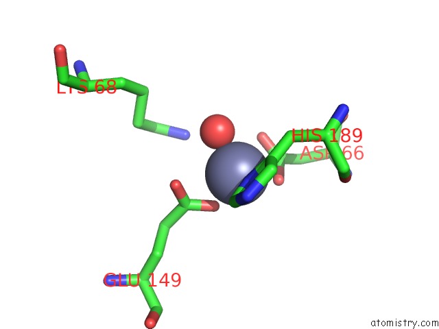
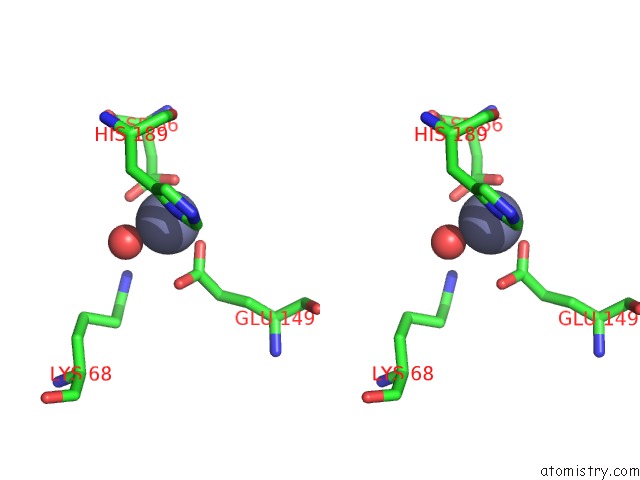
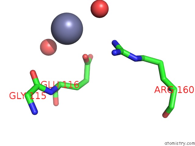
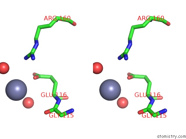
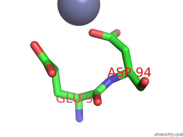
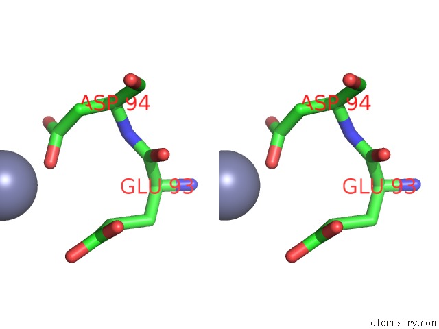
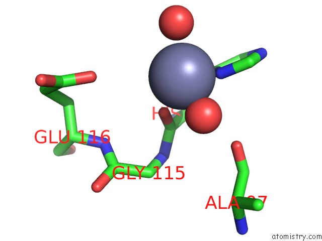
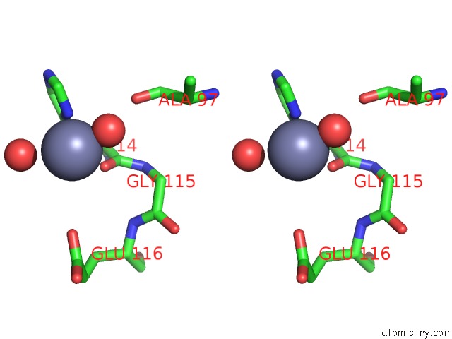
Zinc binding site 1 out of 19 in 3byw
Go back to
Zinc binding site 1 out
of 19 in the Crystal Structure of An Extracellular Domain of Arabinofuranosyltransferase From Corynebacterium Diphtheriae
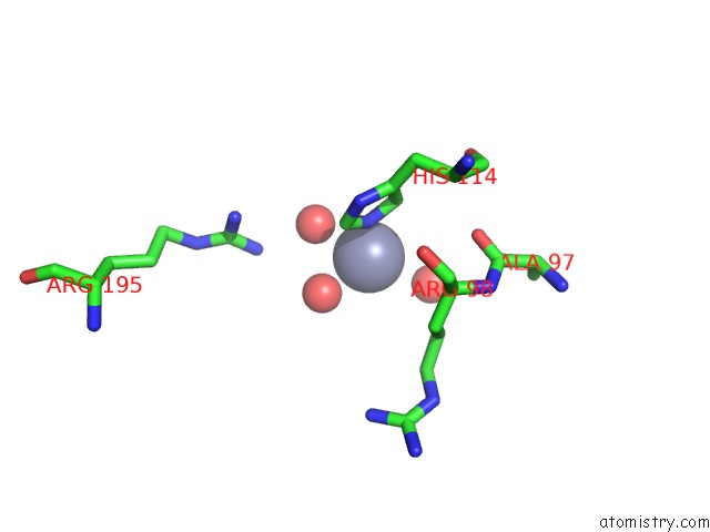
Mono view
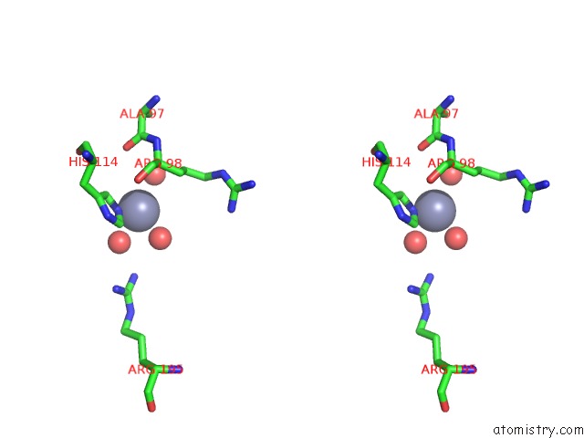
Stereo pair view
Mono view
Stereo pair view
A full contact list of Zinc with other atoms in the Zn binding
site number 1 of Crystal Structure of An Extracellular Domain of Arabinofuranosyltransferase From Corynebacterium Diphtheriae within 5.0Å range:
|
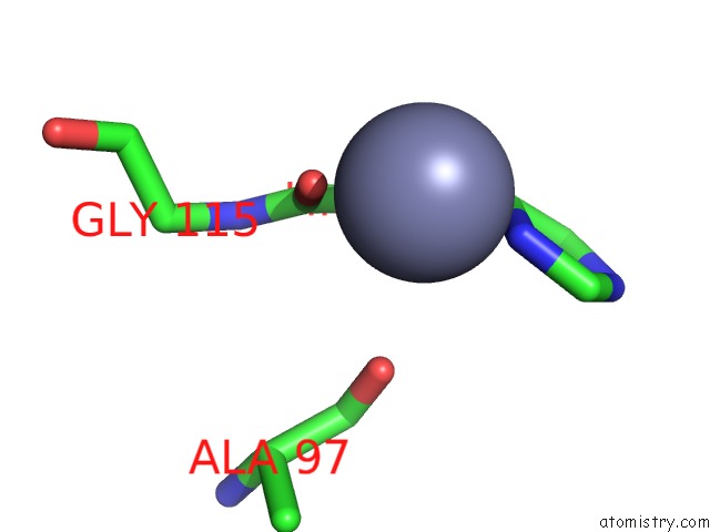
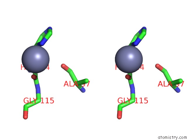
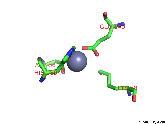
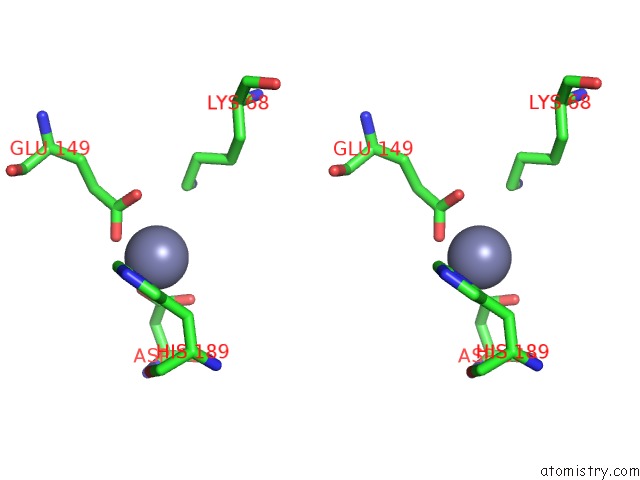
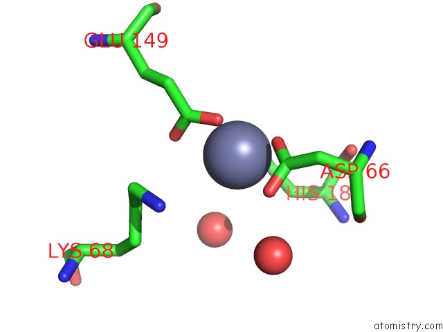
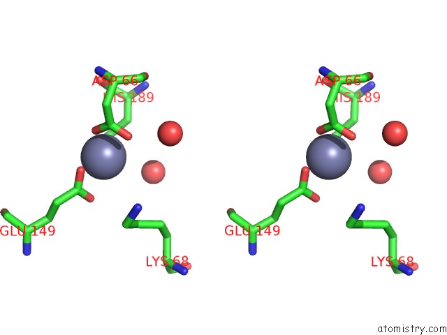
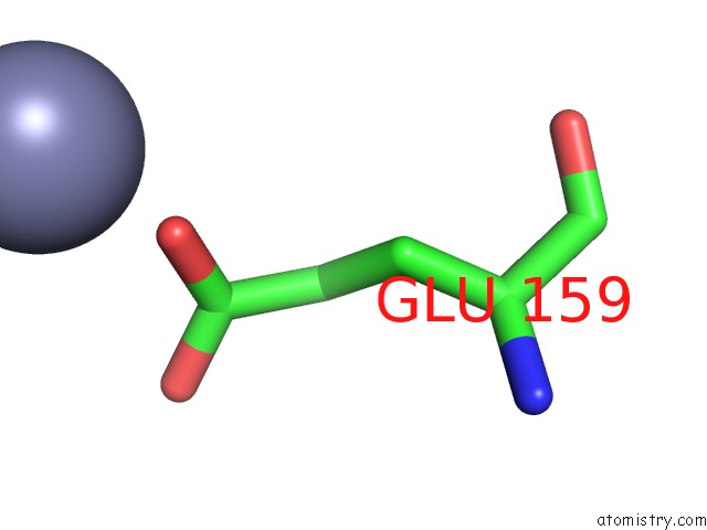
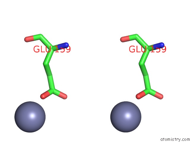
Zinc binding site 2 out of 19 in 3byw
Go back to
Zinc binding site 2 out
of 19 in the Crystal Structure of An Extracellular Domain of Arabinofuranosyltransferase From Corynebacterium Diphtheriae
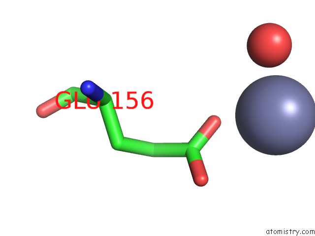
Mono view
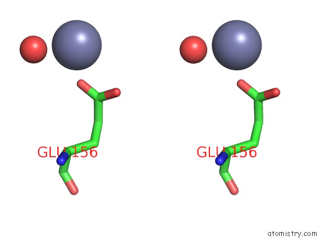
Stereo pair view
Mono view
Stereo pair view
A full contact list of Zinc with other atoms in the Zn binding
site number 2 of Crystal Structure of An Extracellular Domain of Arabinofuranosyltransferase From Corynebacterium Diphtheriae within 5.0Å range:
|
Zinc binding site 3 out of 19 in 3byw
Go back to
Zinc binding site 3 out
of 19 in the Crystal Structure of An Extracellular Domain of Arabinofuranosyltransferase From Corynebacterium Diphtheriae
Mono view
Stereo pair view
Mono view
Stereo pair view
A full contact list of Zinc with other atoms in the Zn binding
site number 3 of Crystal Structure of An Extracellular Domain of Arabinofuranosyltransferase From Corynebacterium Diphtheriae within 5.0Å range:
|
Zinc binding site 4 out of 19 in 3byw
Go back to
Zinc binding site 4 out
of 19 in the Crystal Structure of An Extracellular Domain of Arabinofuranosyltransferase From Corynebacterium Diphtheriae
Mono view
Stereo pair view
Mono view
Stereo pair view
A full contact list of Zinc with other atoms in the Zn binding
site number 4 of Crystal Structure of An Extracellular Domain of Arabinofuranosyltransferase From Corynebacterium Diphtheriae within 5.0Å range:
|
Zinc binding site 5 out of 19 in 3byw
Go back to
Zinc binding site 5 out
of 19 in the Crystal Structure of An Extracellular Domain of Arabinofuranosyltransferase From Corynebacterium Diphtheriae
Mono view
Stereo pair view
Mono view
Stereo pair view
A full contact list of Zinc with other atoms in the Zn binding
site number 5 of Crystal Structure of An Extracellular Domain of Arabinofuranosyltransferase From Corynebacterium Diphtheriae within 5.0Å range:
|
Zinc binding site 6 out of 19 in 3byw
Go back to
Zinc binding site 6 out
of 19 in the Crystal Structure of An Extracellular Domain of Arabinofuranosyltransferase From Corynebacterium Diphtheriae
Mono view
Stereo pair view
Mono view
Stereo pair view
A full contact list of Zinc with other atoms in the Zn binding
site number 6 of Crystal Structure of An Extracellular Domain of Arabinofuranosyltransferase From Corynebacterium Diphtheriae within 5.0Å range:
|
Zinc binding site 7 out of 19 in 3byw
Go back to
Zinc binding site 7 out
of 19 in the Crystal Structure of An Extracellular Domain of Arabinofuranosyltransferase From Corynebacterium Diphtheriae
Mono view
Stereo pair view
Mono view
Stereo pair view
A full contact list of Zinc with other atoms in the Zn binding
site number 7 of Crystal Structure of An Extracellular Domain of Arabinofuranosyltransferase From Corynebacterium Diphtheriae within 5.0Å range:
|
Zinc binding site 8 out of 19 in 3byw
Go back to
Zinc binding site 8 out
of 19 in the Crystal Structure of An Extracellular Domain of Arabinofuranosyltransferase From Corynebacterium Diphtheriae
Mono view
Stereo pair view
Mono view
Stereo pair view
A full contact list of Zinc with other atoms in the Zn binding
site number 8 of Crystal Structure of An Extracellular Domain of Arabinofuranosyltransferase From Corynebacterium Diphtheriae within 5.0Å range:
|
Zinc binding site 9 out of 19 in 3byw
Go back to
Zinc binding site 9 out
of 19 in the Crystal Structure of An Extracellular Domain of Arabinofuranosyltransferase From Corynebacterium Diphtheriae
Mono view
Stereo pair view
Mono view
Stereo pair view
A full contact list of Zinc with other atoms in the Zn binding
site number 9 of Crystal Structure of An Extracellular Domain of Arabinofuranosyltransferase From Corynebacterium Diphtheriae within 5.0Å range:
|
Zinc binding site 10 out of 19 in 3byw
Go back to
Zinc binding site 10 out
of 19 in the Crystal Structure of An Extracellular Domain of Arabinofuranosyltransferase From Corynebacterium Diphtheriae
Mono view
Stereo pair view
Mono view
Stereo pair view
A full contact list of Zinc with other atoms in the Zn binding
site number 10 of Crystal Structure of An Extracellular Domain of Arabinofuranosyltransferase From Corynebacterium Diphtheriae within 5.0Å range:
|
Reference:
K.Tan,
C.Hatzos,
J.Abdullah,
A.Joachimiak.
The Structure of An Extracellular Domain of Arabinofuranosyltransferase From Corynebacterium Diphtheriae. To Be Published.
Page generated: Wed Aug 20 08:06:09 2025
Last articles
Zn in 3SJBZn in 3SIY
Zn in 3SJA
Zn in 3SIP
Zn in 3SHI
Zn in 3SI1
Zn in 3SI2
Zn in 3SI0
Zn in 3SHB
Zn in 3SHZ