Zinc »
PDB 3scl-3sje »
3si1 »
Zinc in PDB 3si1: Structure of Glycosylated Murine Glutaminyl Cyclase
Enzymatic activity of Structure of Glycosylated Murine Glutaminyl Cyclase
All present enzymatic activity of Structure of Glycosylated Murine Glutaminyl Cyclase:
2.3.2.5;
2.3.2.5;
Protein crystallography data
The structure of Structure of Glycosylated Murine Glutaminyl Cyclase, PDB code: 3si1
was solved by
T.Dambe,
D.Carrillo,
C.Parthier,
M.T.Stubbs,
with X-Ray Crystallography technique. A brief refinement statistics is given in the table below:
| Resolution Low / High (Å) | 19.75 / 2.90 |
| Space group | P 21 21 21 |
| Cell size a, b, c (Å), α, β, γ (°) | 43.240, 86.870, 97.160, 90.00, 90.00, 90.00 |
| R / Rfree (%) | 24.5 / 30.1 |
Zinc Binding Sites:
The binding sites of Zinc atom in the Structure of Glycosylated Murine Glutaminyl Cyclase
(pdb code 3si1). This binding sites where shown within
5.0 Angstroms radius around Zinc atom.
In total only one binding site of Zinc was determined in the Structure of Glycosylated Murine Glutaminyl Cyclase, PDB code: 3si1:
In total only one binding site of Zinc was determined in the Structure of Glycosylated Murine Glutaminyl Cyclase, PDB code: 3si1:
Zinc binding site 1 out of 1 in 3si1
Go back to
Zinc binding site 1 out
of 1 in the Structure of Glycosylated Murine Glutaminyl Cyclase
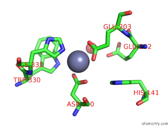
Mono view
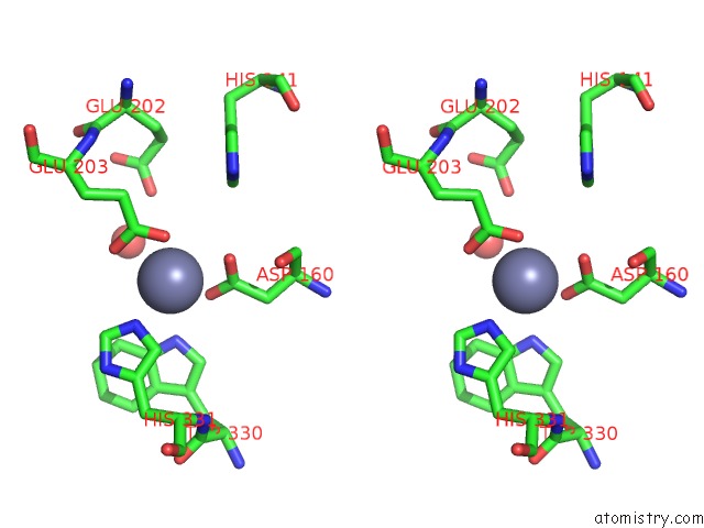
Stereo pair view
Mono view
Stereo pair view
A full contact list of Zinc with other atoms in the Zn binding
site number 1 of Structure of Glycosylated Murine Glutaminyl Cyclase within 5.0Å range:
|
Reference:
D.Ruiz-Carrillo,
B.Koch,
C.Parthier,
M.Wermann,
T.Dambe,
M.Buchholz,
H.H.Ludwig,
U.Heiser,
J.U.Rahfeld,
M.T.Stubbs,
S.Schilling,
H.U.Demuth.
Structures of Glycosylated Mammalian Glutaminyl Cyclases Reveal Conformational Variability Near the Active Center. Biochemistry V. 50 6280 2011.
ISSN: ISSN 0006-2960
PubMed: 21671571
DOI: 10.1021/BI200249H
Page generated: Wed Aug 20 14:03:26 2025
ISSN: ISSN 0006-2960
PubMed: 21671571
DOI: 10.1021/BI200249H
Last articles
Zn in 4JT9Zn in 4JT8
Zn in 4JSZ
Zn in 4JSW
Zn in 4JSR
Zn in 4JSS
Zn in 4JSM
Zn in 4JSL
Zn in 4JSI
Zn in 4JSJ