Zinc »
PDB 3boc-3c4u »
3bvo »
Zinc in PDB 3bvo: Crystal Structure of Human Co-Chaperone Protein Hscb
Protein crystallography data
The structure of Crystal Structure of Human Co-Chaperone Protein Hscb, PDB code: 3bvo
was solved by
E.Bitto,
C.A.Bingman,
J.G.Mccoy,
G.E.Wesenberg,
G.N.Phillips Jr.,
Centerfor Eukaryotic Structural Genomics (Cesg),
with X-Ray Crystallography technique. A brief refinement statistics is given in the table below:
| Resolution Low / High (Å) | 47.75 / 3.00 |
| Space group | P 1 21 1 |
| Cell size a, b, c (Å), α, β, γ (°) | 63.644, 32.581, 114.362, 90.00, 105.24, 90.00 |
| R / Rfree (%) | 23.6 / 28.8 |
Zinc Binding Sites:
The binding sites of Zinc atom in the Crystal Structure of Human Co-Chaperone Protein Hscb
(pdb code 3bvo). This binding sites where shown within
5.0 Angstroms radius around Zinc atom.
In total 2 binding sites of Zinc where determined in the Crystal Structure of Human Co-Chaperone Protein Hscb, PDB code: 3bvo:
Jump to Zinc binding site number: 1; 2;
In total 2 binding sites of Zinc where determined in the Crystal Structure of Human Co-Chaperone Protein Hscb, PDB code: 3bvo:
Jump to Zinc binding site number: 1; 2;
Zinc binding site 1 out of 2 in 3bvo
Go back to
Zinc binding site 1 out
of 2 in the Crystal Structure of Human Co-Chaperone Protein Hscb
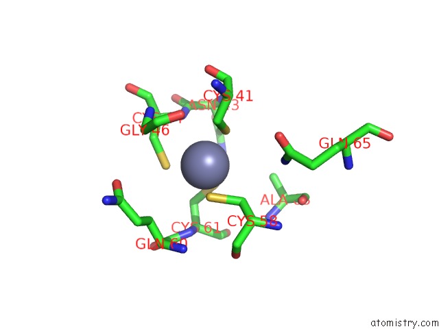
Mono view
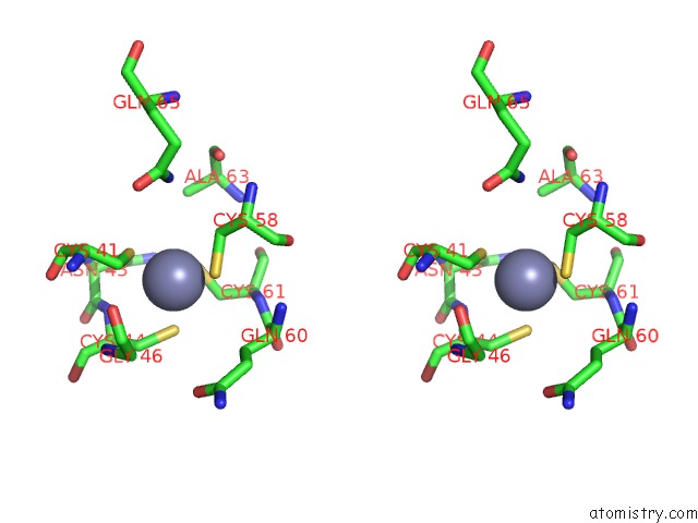
Stereo pair view
Mono view
Stereo pair view
A full contact list of Zinc with other atoms in the Zn binding
site number 1 of Crystal Structure of Human Co-Chaperone Protein Hscb within 5.0Å range:
|
Zinc binding site 2 out of 2 in 3bvo
Go back to
Zinc binding site 2 out
of 2 in the Crystal Structure of Human Co-Chaperone Protein Hscb
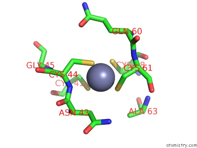
Mono view
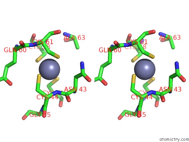
Stereo pair view
Mono view
Stereo pair view
A full contact list of Zinc with other atoms in the Zn binding
site number 2 of Crystal Structure of Human Co-Chaperone Protein Hscb within 5.0Å range:
|
Reference:
E.Bitto,
C.A.Bingman,
L.Bittova,
D.A.Kondrashov,
R.M.Bannen,
B.G.Fox,
J.L.Markley,
G.N.Phillips.
Structure of Human J-Type Co-Chaperone Hscb Reveals A Tetracysteine Metal-Binding Domain. J.Biol.Chem. V. 283 30184 2008.
ISSN: ISSN 0021-9258
PubMed: 18713742
DOI: 10.1074/JBC.M804746200
Page generated: Thu Oct 24 11:38:09 2024
ISSN: ISSN 0021-9258
PubMed: 18713742
DOI: 10.1074/JBC.M804746200
Last articles
Zn in 9J0NZn in 9J0O
Zn in 9J0P
Zn in 9FJX
Zn in 9EKB
Zn in 9C0F
Zn in 9CAH
Zn in 9CH0
Zn in 9CH3
Zn in 9CH1