Zinc »
PDB 2zxw-3ada »
3a9j »
Zinc in PDB 3a9j: Crystal Structure of the Mouse TAB2-Nzf in Complex with LYS63-Linked Di-Ubiquitin
Protein crystallography data
The structure of Crystal Structure of the Mouse TAB2-Nzf in Complex with LYS63-Linked Di-Ubiquitin, PDB code: 3a9j
was solved by
Y.Sato,
A.Yoshikawa,
M.Yamashita,
A.Yamagata,
S.Fukai,
with X-Ray Crystallography technique. A brief refinement statistics is given in the table below:
| Resolution Low / High (Å) | 36.01 / 1.18 |
| Space group | P 21 21 21 |
| Cell size a, b, c (Å), α, β, γ (°) | 30.085, 71.478, 72.029, 90.00, 90.00, 90.00 |
| R / Rfree (%) | 16.4 / 19.1 |
Zinc Binding Sites:
The binding sites of Zinc atom in the Crystal Structure of the Mouse TAB2-Nzf in Complex with LYS63-Linked Di-Ubiquitin
(pdb code 3a9j). This binding sites where shown within
5.0 Angstroms radius around Zinc atom.
In total only one binding site of Zinc was determined in the Crystal Structure of the Mouse TAB2-Nzf in Complex with LYS63-Linked Di-Ubiquitin, PDB code: 3a9j:
In total only one binding site of Zinc was determined in the Crystal Structure of the Mouse TAB2-Nzf in Complex with LYS63-Linked Di-Ubiquitin, PDB code: 3a9j:
Zinc binding site 1 out of 1 in 3a9j
Go back to
Zinc binding site 1 out
of 1 in the Crystal Structure of the Mouse TAB2-Nzf in Complex with LYS63-Linked Di-Ubiquitin
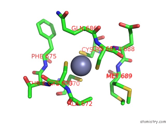
Mono view
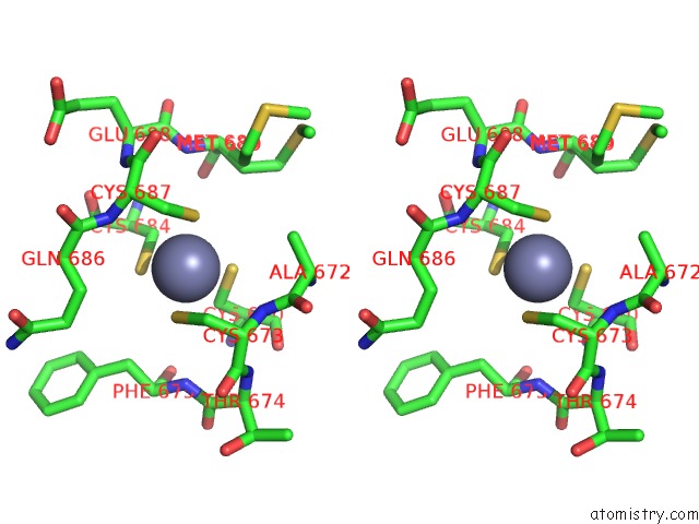
Stereo pair view
Mono view
Stereo pair view
A full contact list of Zinc with other atoms in the Zn binding
site number 1 of Crystal Structure of the Mouse TAB2-Nzf in Complex with LYS63-Linked Di-Ubiquitin within 5.0Å range:
|
Reference:
Y.Sato,
A.Yoshikawa,
M.Yamashita,
A.Yamagata,
S.Fukai.
Structural Basis For Specific Recognition of Lys 63-Linked Polyubiquitin Chains By Nzf Domains of TAB2 and TAB3 Embo J. V. 28 3903 2009.
ISSN: ISSN 0261-4189
PubMed: 19927120
DOI: 10.1038/EMBOJ.2009.345
Page generated: Thu Oct 24 11:07:57 2024
ISSN: ISSN 0261-4189
PubMed: 19927120
DOI: 10.1038/EMBOJ.2009.345
Last articles
Zn in 9J0NZn in 9J0O
Zn in 9J0P
Zn in 9FJX
Zn in 9EKB
Zn in 9C0F
Zn in 9CAH
Zn in 9CH0
Zn in 9CH3
Zn in 9CH1