Zinc »
PDB 2zze-3adr »
3a43 »
Zinc in PDB 3a43: Crystal Structure of Hypa
Protein crystallography data
The structure of Crystal Structure of Hypa, PDB code: 3a43
was solved by
S.Watanabe,
T.Arai,
R.Matsumi,
H.Aromi,
T.Imanaka,
K.Miki,
with X-Ray Crystallography technique. A brief refinement statistics is given in the table below:
| Resolution Low / High (Å) | 41.97 / 2.30 |
| Space group | H 3 2 |
| Cell size a, b, c (Å), α, β, γ (°) | 83.933, 83.933, 365.006, 90.00, 90.00, 120.00 |
| R / Rfree (%) | 21.5 / 23.9 |
Zinc Binding Sites:
The binding sites of Zinc atom in the Crystal Structure of Hypa
(pdb code 3a43). This binding sites where shown within
5.0 Angstroms radius around Zinc atom.
In total 2 binding sites of Zinc where determined in the Crystal Structure of Hypa, PDB code: 3a43:
Jump to Zinc binding site number: 1; 2;
In total 2 binding sites of Zinc where determined in the Crystal Structure of Hypa, PDB code: 3a43:
Jump to Zinc binding site number: 1; 2;
Zinc binding site 1 out of 2 in 3a43
Go back to
Zinc binding site 1 out
of 2 in the Crystal Structure of Hypa
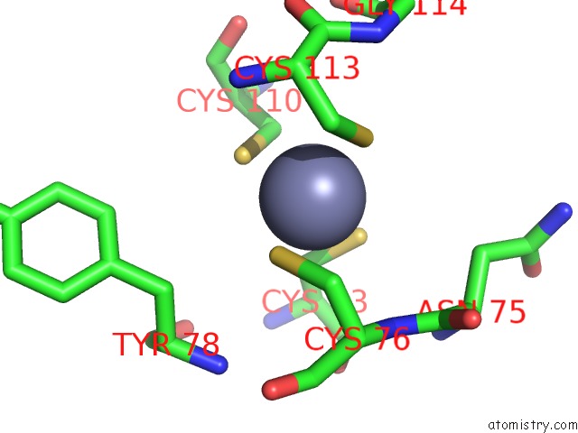
Mono view
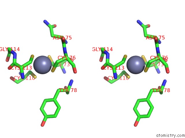
Stereo pair view
Mono view
Stereo pair view
A full contact list of Zinc with other atoms in the Zn binding
site number 1 of Crystal Structure of Hypa within 5.0Å range:
|
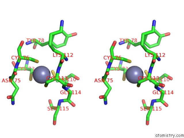
Zinc binding site 2 out of 2 in 3a43
Go back to
Zinc binding site 2 out
of 2 in the Crystal Structure of Hypa
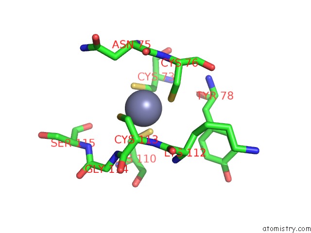
Mono view
Stereo pair view
Mono view
Stereo pair view
A full contact list of Zinc with other atoms in the Zn binding
site number 2 of Crystal Structure of Hypa within 5.0Å range:
|
Reference:
S.Watanabe,
T.Arai,
R.Matsumi,
H.Atomi,
T.Imanaka,
K.Miki.
Crystal Structure of Hypa, A Nickel-Binding Metallochaperone For [Nife] Hydrogenase Maturation. J.Mol.Biol. V. 394 448 2009.
ISSN: ISSN 0022-2836
PubMed: 19769985
DOI: 10.1016/J.JMB.2009.09.030
Page generated: Wed Aug 20 07:39:32 2025
ISSN: ISSN 0022-2836
PubMed: 19769985
DOI: 10.1016/J.JMB.2009.09.030
Last articles
Zn in 3KRVZn in 3KR8
Zn in 3KR7
Zn in 3KQX
Zn in 3KM2
Zn in 3KL9
Zn in 3KQI
Zn in 3KQ6
Zn in 3KNX
Zn in 3KNV