Zinc »
PDB 3kew-3kr8 »
3kqx »
Zinc in PDB 3kqx: Structure of A Protease 1
Enzymatic activity of Structure of A Protease 1
All present enzymatic activity of Structure of A Protease 1:
3.4.11.1;
3.4.11.1;
Protein crystallography data
The structure of Structure of A Protease 1, PDB code: 3kqx
was solved by
S.Mcgowan,
J.C.Whisstock,
with X-Ray Crystallography technique. A brief refinement statistics is given in the table below:
| Resolution Low / High (Å) | 56.08 / 2.01 |
| Space group | P 21 21 21 |
| Cell size a, b, c (Å), α, β, γ (°) | 173.404, 176.809, 224.246, 90.00, 90.00, 90.00 |
| R / Rfree (%) | 18 / 23.2 |
Zinc Binding Sites:
Pages:
>>> Page 1 <<< Page 2, Binding sites: 11 - 12;Binding sites:
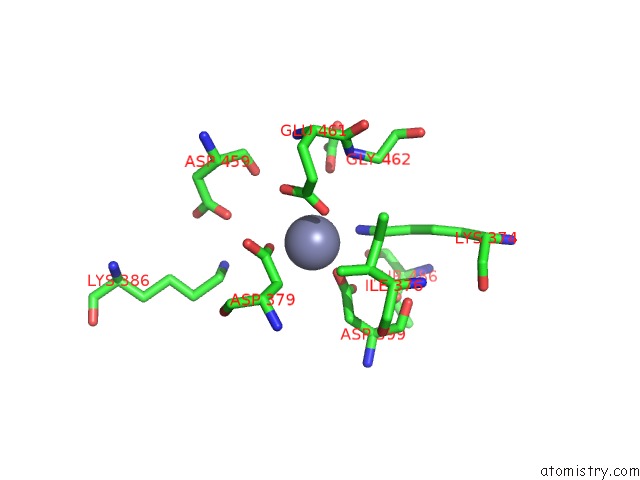
The binding sites of Zinc atom in the Structure of A Protease 1 (pdb code 3kqx). This binding sites where shown within 5.0 Angstroms radius around Zinc atom.In total 12 binding sites of Zinc where determined in the Structure of A Protease 1, PDB code: 3kqx:
Jump to Zinc binding site number: 1; 2; 3; 4; 5; 6; 7; 8; 9; 10;
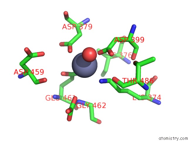
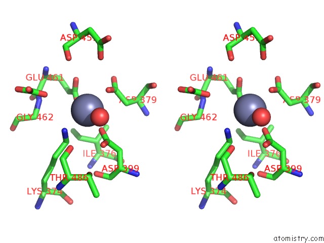
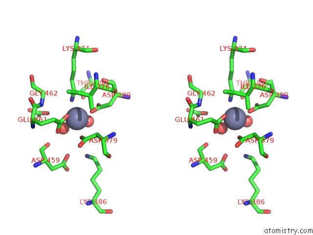
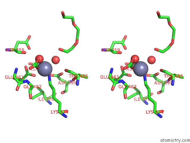
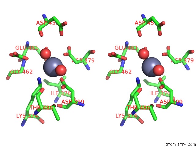
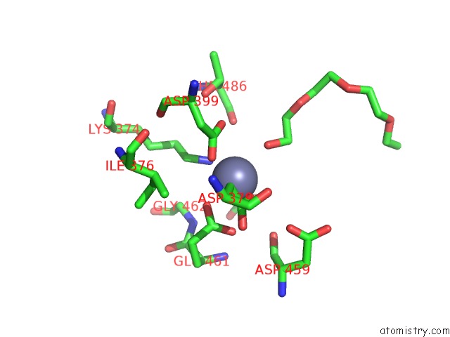
Zinc binding site 1 out of 12 in 3kqx
Go back to
Zinc binding site 1 out
of 12 in the Structure of A Protease 1

Mono view
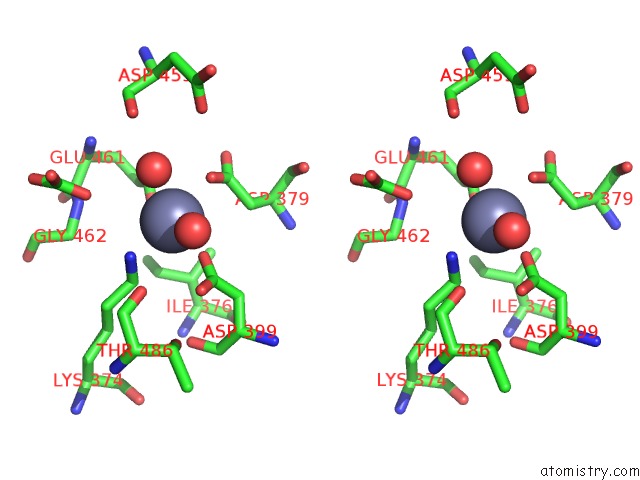
Stereo pair view
Mono view
Stereo pair view
A full contact list of Zinc with other atoms in the Zn binding
site number 1 of Structure of A Protease 1 within 5.0Å range:
|
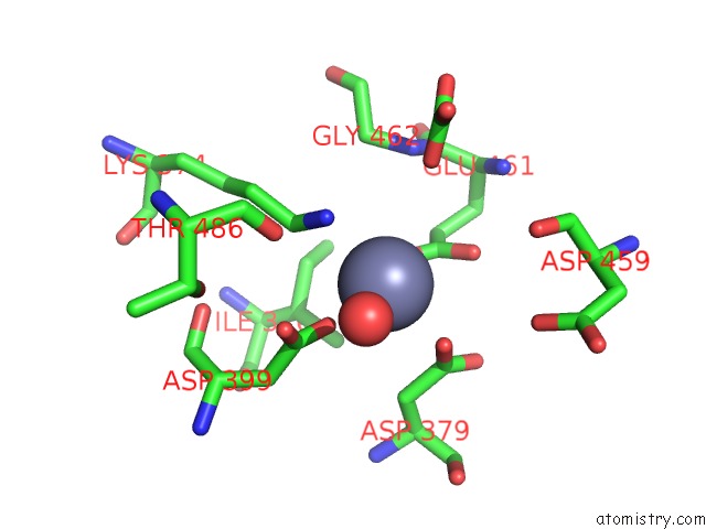
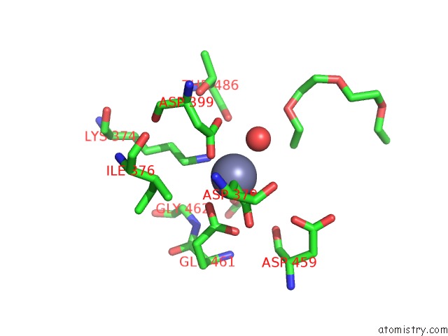
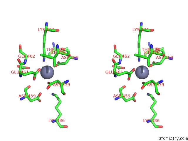
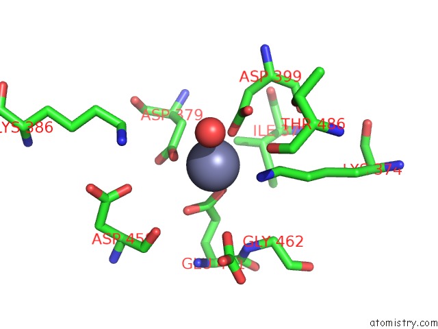
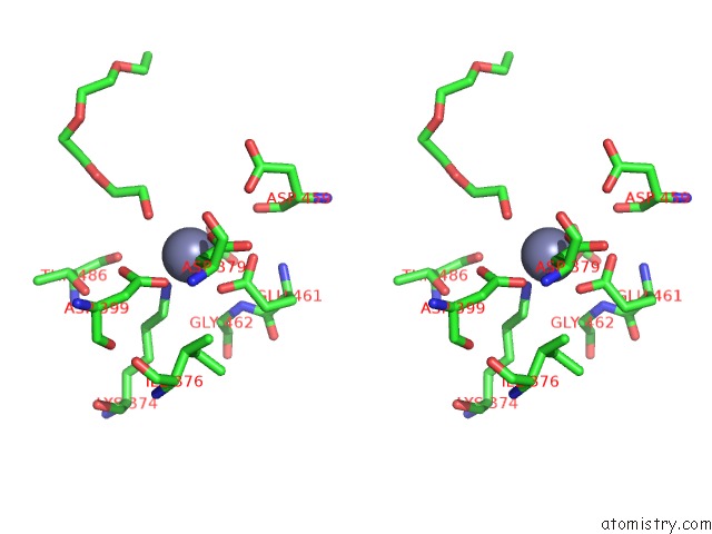
Zinc binding site 2 out of 12 in 3kqx
Go back to
Zinc binding site 2 out
of 12 in the Structure of A Protease 1
Mono view
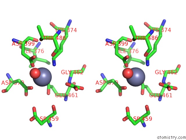
Stereo pair view
Mono view
Stereo pair view
A full contact list of Zinc with other atoms in the Zn binding
site number 2 of Structure of A Protease 1 within 5.0Å range:
|
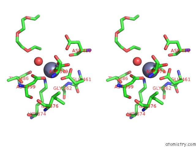
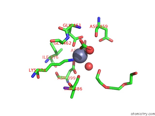
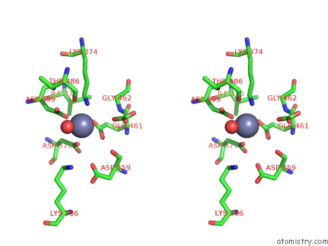
Zinc binding site 3 out of 12 in 3kqx
Go back to
Zinc binding site 3 out
of 12 in the Structure of A Protease 1
Mono view
Stereo pair view
Mono view
Stereo pair view
A full contact list of Zinc with other atoms in the Zn binding
site number 3 of Structure of A Protease 1 within 5.0Å range:
|
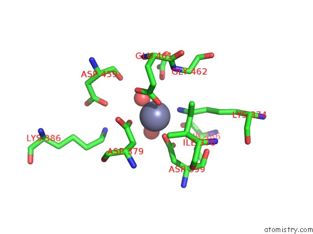
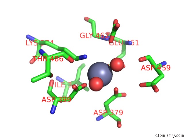
Zinc binding site 4 out of 12 in 3kqx
Go back to
Zinc binding site 4 out
of 12 in the Structure of A Protease 1
Mono view
Stereo pair view
Mono view
Stereo pair view
A full contact list of Zinc with other atoms in the Zn binding
site number 4 of Structure of A Protease 1 within 5.0Å range:
|
Zinc binding site 5 out of 12 in 3kqx
Go back to
Zinc binding site 5 out
of 12 in the Structure of A Protease 1
Mono view
Stereo pair view
Mono view
Stereo pair view
A full contact list of Zinc with other atoms in the Zn binding
site number 5 of Structure of A Protease 1 within 5.0Å range:
|
Zinc binding site 6 out of 12 in 3kqx
Go back to
Zinc binding site 6 out
of 12 in the Structure of A Protease 1
Mono view
Stereo pair view
Mono view
Stereo pair view
A full contact list of Zinc with other atoms in the Zn binding
site number 6 of Structure of A Protease 1 within 5.0Å range:
|
Zinc binding site 7 out of 12 in 3kqx
Go back to
Zinc binding site 7 out
of 12 in the Structure of A Protease 1
Mono view
Stereo pair view
Mono view
Stereo pair view
A full contact list of Zinc with other atoms in the Zn binding
site number 7 of Structure of A Protease 1 within 5.0Å range:
|
Zinc binding site 8 out of 12 in 3kqx
Go back to
Zinc binding site 8 out
of 12 in the Structure of A Protease 1
Mono view
Stereo pair view
Mono view
Stereo pair view
A full contact list of Zinc with other atoms in the Zn binding
site number 8 of Structure of A Protease 1 within 5.0Å range:
|
Zinc binding site 9 out of 12 in 3kqx
Go back to
Zinc binding site 9 out
of 12 in the Structure of A Protease 1
Mono view
Stereo pair view
Mono view
Stereo pair view
A full contact list of Zinc with other atoms in the Zn binding
site number 9 of Structure of A Protease 1 within 5.0Å range:
|
Zinc binding site 10 out of 12 in 3kqx
Go back to
Zinc binding site 10 out
of 12 in the Structure of A Protease 1
Mono view
Stereo pair view
Mono view
Stereo pair view
A full contact list of Zinc with other atoms in the Zn binding
site number 10 of Structure of A Protease 1 within 5.0Å range:
|
Reference:
S.Mcgowan,
C.A.Oellig,
W.A.Birru,
T.T.Caradoc-Davies,
C.M.Stack,
J.Lowther,
T.Skinner-Adams,
A.Mucha,
P.Kafarski,
J.Grembecka,
K.R.Trenholme,
A.M.Buckle,
D.L.Gardiner,
J.P.Dalton,
J.C.Whisstock.
Structure of the Plasmodium Falciparum M17 Aminopeptidase and Significance For the Design of Drugs Targeting the Neutral Exopeptidases Proc.Natl.Acad.Sci.Usa V. 107 2449 2010.
ISSN: ISSN 0027-8424
PubMed: 20133789
DOI: 10.1073/PNAS.0911813107
Page generated: Sat Oct 26 07:59:04 2024
ISSN: ISSN 0027-8424
PubMed: 20133789
DOI: 10.1073/PNAS.0911813107
Last articles
Mg in 1SSQMg in 1SS4
Mg in 1SR6
Mg in 1SO6
Mg in 1SQK
Mg in 1SMY
Mg in 1SO2
Mg in 1SO5
Mg in 1SO4
Mg in 1SO3