Zinc »
PDB 2x91-2xjz »
2xhm »
Zinc in PDB 2xhm: Crystal Structure of Ance-K26 Complex
Enzymatic activity of Crystal Structure of Ance-K26 Complex
All present enzymatic activity of Crystal Structure of Ance-K26 Complex:
3.4.15.1;
3.4.15.1;
Protein crystallography data
The structure of Crystal Structure of Ance-K26 Complex, PDB code: 2xhm
was solved by
M.Akif,
I.Ntai,
E.D.Sturrock,
R.E.Isaac,
B.O.Bachmann,
K.R.Acharya,
with X-Ray Crystallography technique. A brief refinement statistics is given in the table below:
| Resolution Low / High (Å) | 32.64 / 1.96 |
| Space group | H 3 |
| Cell size a, b, c (Å), α, β, γ (°) | 172.707, 172.707, 101.696, 90.00, 90.00, 120.00 |
| R / Rfree (%) | 19.403 / 21.461 |
Zinc Binding Sites:
The binding sites of Zinc atom in the Crystal Structure of Ance-K26 Complex
(pdb code 2xhm). This binding sites where shown within
5.0 Angstroms radius around Zinc atom.
In total only one binding site of Zinc was determined in the Crystal Structure of Ance-K26 Complex, PDB code: 2xhm:
In total only one binding site of Zinc was determined in the Crystal Structure of Ance-K26 Complex, PDB code: 2xhm:
Zinc binding site 1 out of 1 in 2xhm
Go back to
Zinc binding site 1 out
of 1 in the Crystal Structure of Ance-K26 Complex
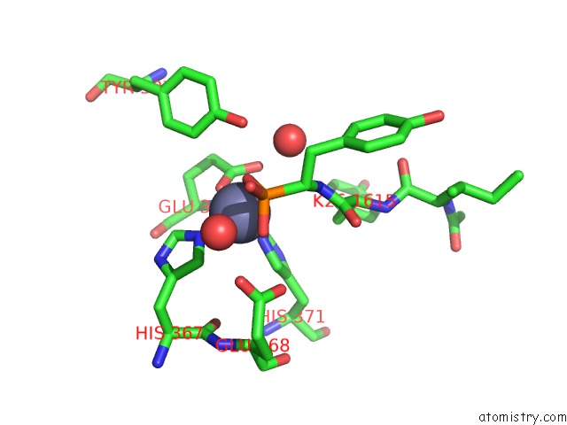
Mono view
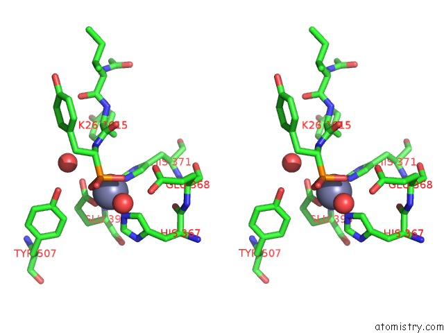
Stereo pair view
Mono view
Stereo pair view
A full contact list of Zinc with other atoms in the Zn binding
site number 1 of Crystal Structure of Ance-K26 Complex within 5.0Å range:
|
Reference:
M.Akif,
I.Ntai,
E.D.Sturrock,
R.E.Isaac,
B.O.Bachmann,
K.R.Acharya.
Crystal Structure of A Phosphonotripeptide K-26 in Complex with Angiotensin Converting Enzyme Homologue (Ance) From Drosophila Melanogaster. Biochem.Biophys.Res.Commun. V. 398 532 2010.
ISSN: ISSN 0006-291X
PubMed: 20599761
DOI: 10.1016/J.BBRC.2010.06.113
Page generated: Thu Oct 17 05:18:56 2024
ISSN: ISSN 0006-291X
PubMed: 20599761
DOI: 10.1016/J.BBRC.2010.06.113
Last articles
Zn in 9J0NZn in 9J0O
Zn in 9J0P
Zn in 9FJX
Zn in 9EKB
Zn in 9C0F
Zn in 9CAH
Zn in 9CH0
Zn in 9CH3
Zn in 9CH1