Zinc »
PDB 2x91-2xjz »
2xbq »
Zinc in PDB 2xbq: Crystal Structure of Reduced Schistosoma Mansoni Thioredoxin Pre-Protein at 1.7 Angstrom
Enzymatic activity of Crystal Structure of Reduced Schistosoma Mansoni Thioredoxin Pre-Protein at 1.7 Angstrom
All present enzymatic activity of Crystal Structure of Reduced Schistosoma Mansoni Thioredoxin Pre-Protein at 1.7 Angstrom:
1.8.1.8;
1.8.1.8;
Protein crystallography data
The structure of Crystal Structure of Reduced Schistosoma Mansoni Thioredoxin Pre-Protein at 1.7 Angstrom, PDB code: 2xbq
was solved by
G.Boumis,
A.E.Miele,
D.Dimastrogiovanni,
F.Angelucci,
A.Bellelli,
with X-Ray Crystallography technique. A brief refinement statistics is given in the table below:
| Resolution Low / High (Å) | 53.84 / 1.67 |
| Space group | P 32 |
| Cell size a, b, c (Å), α, β, γ (°) | 62.143, 62.143, 58.292, 90.00, 90.00, 120.00 |
| R / Rfree (%) | 21.468 / 24.184 |
Zinc Binding Sites:
The binding sites of Zinc atom in the Crystal Structure of Reduced Schistosoma Mansoni Thioredoxin Pre-Protein at 1.7 Angstrom
(pdb code 2xbq). This binding sites where shown within
5.0 Angstroms radius around Zinc atom.
In total 2 binding sites of Zinc where determined in the Crystal Structure of Reduced Schistosoma Mansoni Thioredoxin Pre-Protein at 1.7 Angstrom, PDB code: 2xbq:
Jump to Zinc binding site number: 1; 2;
In total 2 binding sites of Zinc where determined in the Crystal Structure of Reduced Schistosoma Mansoni Thioredoxin Pre-Protein at 1.7 Angstrom, PDB code: 2xbq:
Jump to Zinc binding site number: 1; 2;
Zinc binding site 1 out of 2 in 2xbq
Go back to
Zinc binding site 1 out
of 2 in the Crystal Structure of Reduced Schistosoma Mansoni Thioredoxin Pre-Protein at 1.7 Angstrom
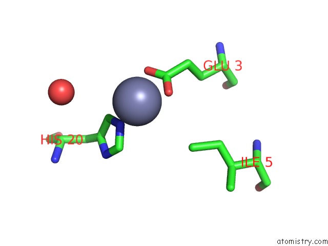
Mono view
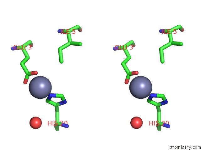
Stereo pair view
Mono view
Stereo pair view
A full contact list of Zinc with other atoms in the Zn binding
site number 1 of Crystal Structure of Reduced Schistosoma Mansoni Thioredoxin Pre-Protein at 1.7 Angstrom within 5.0Å range:
|
Zinc binding site 2 out of 2 in 2xbq
Go back to
Zinc binding site 2 out
of 2 in the Crystal Structure of Reduced Schistosoma Mansoni Thioredoxin Pre-Protein at 1.7 Angstrom
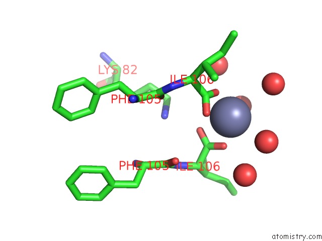
Mono view

Stereo pair view
Mono view
Stereo pair view
A full contact list of Zinc with other atoms in the Zn binding
site number 2 of Crystal Structure of Reduced Schistosoma Mansoni Thioredoxin Pre-Protein at 1.7 Angstrom within 5.0Å range:
|
Reference:
G.Boumis,
F.Angelucci,
A.Bellelli,
M.Brunori,
D.Dimastrogiovanni,
A.E.Miele.
Structural and Functional Characterization of Schistosoma Mansoni Thioredoxin. Protein Sci. V. 20 1069 2011.
ISSN: ISSN 0961-8368
PubMed: 21465612
DOI: 10.1002/PRO.634
Page generated: Thu Oct 17 05:16:10 2024
ISSN: ISSN 0961-8368
PubMed: 21465612
DOI: 10.1002/PRO.634
Last articles
Zn in 9J0NZn in 9J0O
Zn in 9J0P
Zn in 9FJX
Zn in 9EKB
Zn in 9C0F
Zn in 9CAH
Zn in 9CH0
Zn in 9CH3
Zn in 9CH1