Zinc »
PDB 2wwz-2x90 »
2wz5 »
Zinc in PDB 2wz5: L38V SOD1 Mutant Complexed with L-Methionine.
Enzymatic activity of L38V SOD1 Mutant Complexed with L-Methionine.
All present enzymatic activity of L38V SOD1 Mutant Complexed with L-Methionine.:
1.15.1.1;
1.15.1.1;
Protein crystallography data
The structure of L38V SOD1 Mutant Complexed with L-Methionine., PDB code: 2wz5
was solved by
S.V.Antonyuk,
R.W.Strange,
S.S.Hasnain,
with X-Ray Crystallography technique. A brief refinement statistics is given in the table below:
| Resolution Low / High (Å) | 24.00 / 1.50 |
| Space group | P 1 21 1 |
| Cell size a, b, c (Å), α, β, γ (°) | 38.996, 68.025, 50.633, 90.00, 105.88, 90.00 |
| R / Rfree (%) | 19.2 / 25.5 |
Other elements in 2wz5:
The structure of L38V SOD1 Mutant Complexed with L-Methionine. also contains other interesting chemical elements:
| Copper | (Cu) | 3 atoms |
Zinc Binding Sites:
The binding sites of Zinc atom in the L38V SOD1 Mutant Complexed with L-Methionine.
(pdb code 2wz5). This binding sites where shown within
5.0 Angstroms radius around Zinc atom.
In total 3 binding sites of Zinc where determined in the L38V SOD1 Mutant Complexed with L-Methionine., PDB code: 2wz5:
Jump to Zinc binding site number: 1; 2; 3;
In total 3 binding sites of Zinc where determined in the L38V SOD1 Mutant Complexed with L-Methionine., PDB code: 2wz5:
Jump to Zinc binding site number: 1; 2; 3;
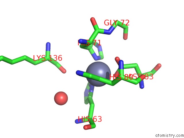
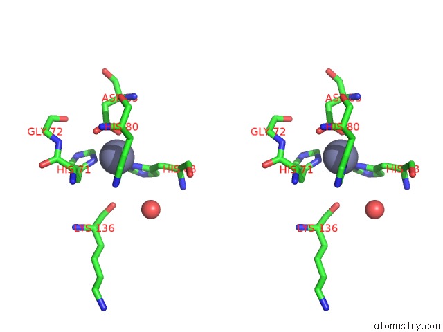
Zinc binding site 1 out of 3 in 2wz5
Go back to
Zinc binding site 1 out
of 3 in the L38V SOD1 Mutant Complexed with L-Methionine.
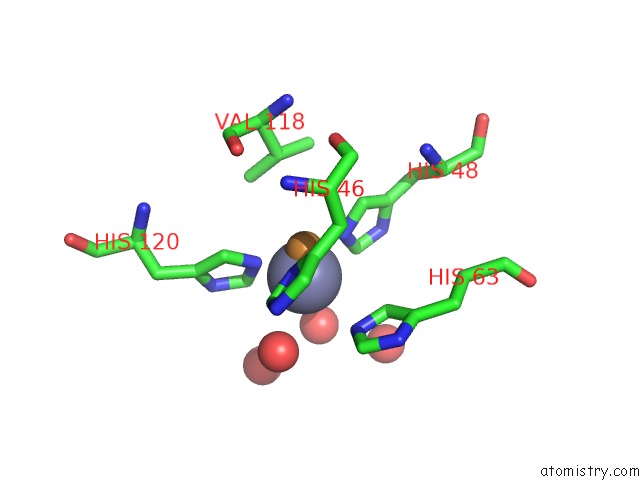
Mono view
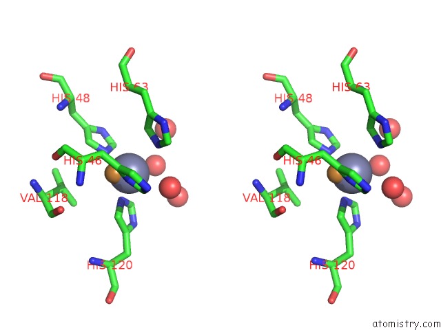
Stereo pair view
Mono view
Stereo pair view
A full contact list of Zinc with other atoms in the Zn binding
site number 1 of L38V SOD1 Mutant Complexed with L-Methionine. within 5.0Å range:
|
Zinc binding site 2 out of 3 in 2wz5
Go back to
Zinc binding site 2 out
of 3 in the L38V SOD1 Mutant Complexed with L-Methionine.
Mono view
Stereo pair view
Mono view
Stereo pair view
A full contact list of Zinc with other atoms in the Zn binding
site number 2 of L38V SOD1 Mutant Complexed with L-Methionine. within 5.0Å range:
|
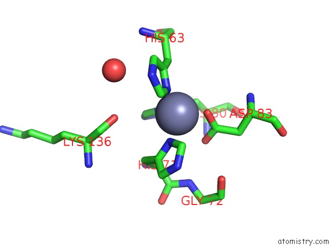
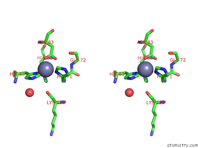
Zinc binding site 3 out of 3 in 2wz5
Go back to
Zinc binding site 3 out
of 3 in the L38V SOD1 Mutant Complexed with L-Methionine.
Mono view
Stereo pair view
Mono view
Stereo pair view
A full contact list of Zinc with other atoms in the Zn binding
site number 3 of L38V SOD1 Mutant Complexed with L-Methionine. within 5.0Å range:
|
Reference:
S.Antonyuk,
R.W.Strange,
S.S.Hasnain.
Structural Discovery of Small Molecule Binding Sites in Cu-Zn Human Superoxide Dismutase Familial Amyotrophic Lateral Sclerosis Mutants Provides Insights For Lead Optimization. J.Med.Chem. V. 53 1402 2010.
ISSN: ISSN 0022-2623
PubMed: 20067275
DOI: 10.1021/JM9017948
Page generated: Thu Oct 17 05:05:44 2024
ISSN: ISSN 0022-2623
PubMed: 20067275
DOI: 10.1021/JM9017948
Last articles
Zn in 9J0NZn in 9J0O
Zn in 9J0P
Zn in 9FJX
Zn in 9EKB
Zn in 9C0F
Zn in 9CAH
Zn in 9CH0
Zn in 9CH3
Zn in 9CH1