Zinc »
PDB 2vr6-2w15 »
2vr7 »
Zinc in PDB 2vr7: Crystal Structure of G85R Als Mutant of Human Cu,Zn Superoxide Dismutase (Cuznsod) at 1.58 A Resolution
Enzymatic activity of Crystal Structure of G85R Als Mutant of Human Cu,Zn Superoxide Dismutase (Cuznsod) at 1.58 A Resolution
All present enzymatic activity of Crystal Structure of G85R Als Mutant of Human Cu,Zn Superoxide Dismutase (Cuznsod) at 1.58 A Resolution:
1.15.1.1;
1.15.1.1;
Protein crystallography data
The structure of Crystal Structure of G85R Als Mutant of Human Cu,Zn Superoxide Dismutase (Cuznsod) at 1.58 A Resolution, PDB code: 2vr7
was solved by
S.Antonyuk,
X.Cao,
S.V.Seetharaman,
L.J.Whitson,
A.B.Taylor,
S.P.Holloway,
R.W.Strange,
P.A.Doucette,
A.Tiwari,
L.J.Hayward,
S.Padua,
J.A.Cohlberg,
J.Selverstone Valentine,
S.S.Hasnain,
P.J.Hart,
with X-Ray Crystallography technique. A brief refinement statistics is given in the table below:
| Resolution Low / High (Å) | 20.00 / 1.58 |
| Space group | P 1 21 1 |
| Cell size a, b, c (Å), α, β, γ (°) | 36.768, 56.435, 75.028, 90.00, 102.98, 90.00 |
| R / Rfree (%) | 13.5 / 17.5 |
Other elements in 2vr7:
The structure of Crystal Structure of G85R Als Mutant of Human Cu,Zn Superoxide Dismutase (Cuznsod) at 1.58 A Resolution also contains other interesting chemical elements:
| Copper | (Cu) | 2 atoms |
Zinc Binding Sites:
The binding sites of Zinc atom in the Crystal Structure of G85R Als Mutant of Human Cu,Zn Superoxide Dismutase (Cuznsod) at 1.58 A Resolution
(pdb code 2vr7). This binding sites where shown within
5.0 Angstroms radius around Zinc atom.
In total 6 binding sites of Zinc where determined in the Crystal Structure of G85R Als Mutant of Human Cu,Zn Superoxide Dismutase (Cuznsod) at 1.58 A Resolution, PDB code: 2vr7:
Jump to Zinc binding site number: 1; 2; 3; 4; 5; 6;
In total 6 binding sites of Zinc where determined in the Crystal Structure of G85R Als Mutant of Human Cu,Zn Superoxide Dismutase (Cuznsod) at 1.58 A Resolution, PDB code: 2vr7:
Jump to Zinc binding site number: 1; 2; 3; 4; 5; 6;
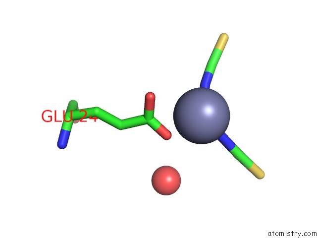
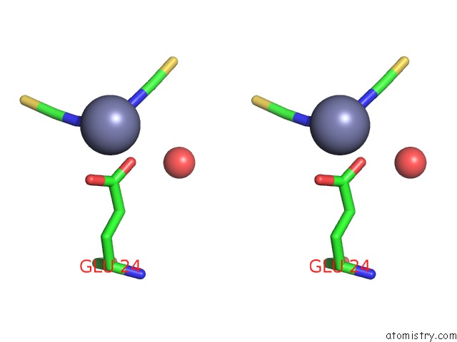
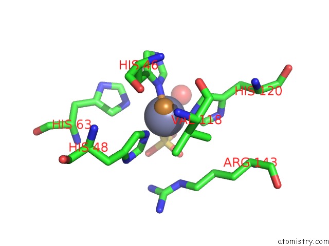
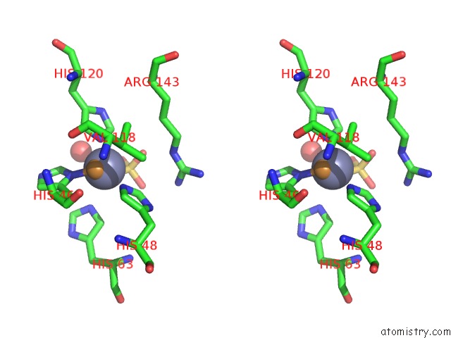
Zinc binding site 1 out of 6 in 2vr7
Go back to
Zinc binding site 1 out
of 6 in the Crystal Structure of G85R Als Mutant of Human Cu,Zn Superoxide Dismutase (Cuznsod) at 1.58 A Resolution
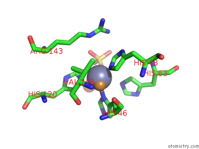
Mono view
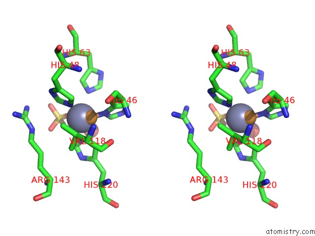
Stereo pair view
Mono view
Stereo pair view
A full contact list of Zinc with other atoms in the Zn binding
site number 1 of Crystal Structure of G85R Als Mutant of Human Cu,Zn Superoxide Dismutase (Cuznsod) at 1.58 A Resolution within 5.0Å range:
|
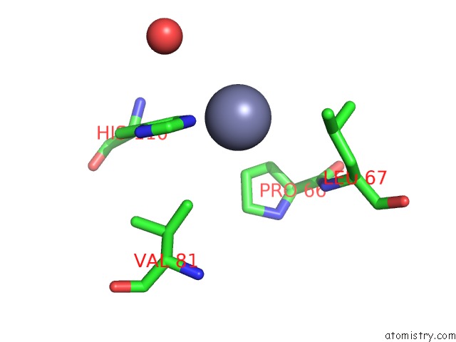
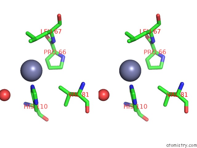
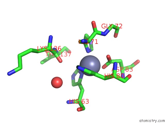
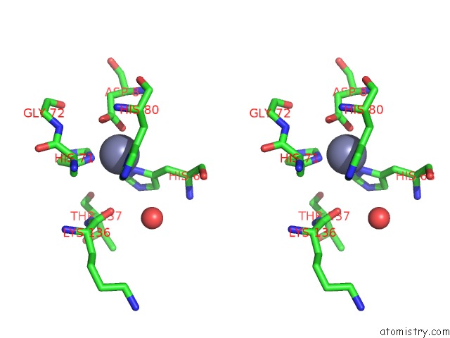
Zinc binding site 2 out of 6 in 2vr7
Go back to
Zinc binding site 2 out
of 6 in the Crystal Structure of G85R Als Mutant of Human Cu,Zn Superoxide Dismutase (Cuznsod) at 1.58 A Resolution
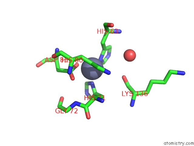
Mono view
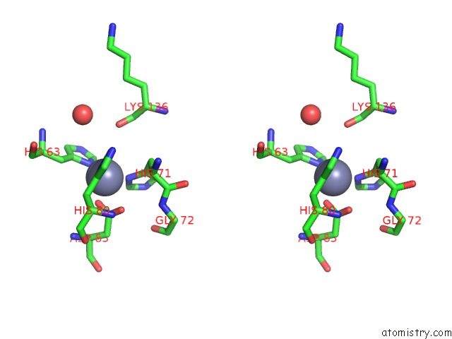
Stereo pair view
Mono view
Stereo pair view
A full contact list of Zinc with other atoms in the Zn binding
site number 2 of Crystal Structure of G85R Als Mutant of Human Cu,Zn Superoxide Dismutase (Cuznsod) at 1.58 A Resolution within 5.0Å range:
|
Zinc binding site 3 out of 6 in 2vr7
Go back to
Zinc binding site 3 out
of 6 in the Crystal Structure of G85R Als Mutant of Human Cu,Zn Superoxide Dismutase (Cuznsod) at 1.58 A Resolution
Mono view
Stereo pair view
Mono view
Stereo pair view
A full contact list of Zinc with other atoms in the Zn binding
site number 3 of Crystal Structure of G85R Als Mutant of Human Cu,Zn Superoxide Dismutase (Cuznsod) at 1.58 A Resolution within 5.0Å range:
|
Zinc binding site 4 out of 6 in 2vr7
Go back to
Zinc binding site 4 out
of 6 in the Crystal Structure of G85R Als Mutant of Human Cu,Zn Superoxide Dismutase (Cuznsod) at 1.58 A Resolution
Mono view
Stereo pair view
Mono view
Stereo pair view
A full contact list of Zinc with other atoms in the Zn binding
site number 4 of Crystal Structure of G85R Als Mutant of Human Cu,Zn Superoxide Dismutase (Cuznsod) at 1.58 A Resolution within 5.0Å range:
|
Zinc binding site 5 out of 6 in 2vr7
Go back to
Zinc binding site 5 out
of 6 in the Crystal Structure of G85R Als Mutant of Human Cu,Zn Superoxide Dismutase (Cuznsod) at 1.58 A Resolution
Mono view
Stereo pair view
Mono view
Stereo pair view
A full contact list of Zinc with other atoms in the Zn binding
site number 5 of Crystal Structure of G85R Als Mutant of Human Cu,Zn Superoxide Dismutase (Cuznsod) at 1.58 A Resolution within 5.0Å range:
|
Zinc binding site 6 out of 6 in 2vr7
Go back to
Zinc binding site 6 out
of 6 in the Crystal Structure of G85R Als Mutant of Human Cu,Zn Superoxide Dismutase (Cuznsod) at 1.58 A Resolution
Mono view
Stereo pair view
Mono view
Stereo pair view
A full contact list of Zinc with other atoms in the Zn binding
site number 6 of Crystal Structure of G85R Als Mutant of Human Cu,Zn Superoxide Dismutase (Cuznsod) at 1.58 A Resolution within 5.0Å range:
|
Reference:
X.Cao,
S.Antonyuk,
S.V.Seetharaman,
L.J.Whitson,
A.B.Taylor,
S.P.Holloway,
R.W.Strange,
P.A.Doucette,
J.S.Valentine,
A.Tiwari,
L.J.Hayward,
S.Padua,
J.A.Cohlberg,
S.S.Hasnain,
P.J.Hart.
Structures of the G85R Variant of SOD1 in Familial Amyotrophic Lateral Sclerosis. J.Biol.Chem. V. 283 16169 2008.
ISSN: ISSN 0021-9258
PubMed: 18378676
DOI: 10.1074/JBC.M801522200
Page generated: Thu Oct 17 04:31:01 2024
ISSN: ISSN 0021-9258
PubMed: 18378676
DOI: 10.1074/JBC.M801522200
Last articles
Zn in 9J0NZn in 9J0O
Zn in 9J0P
Zn in 9FJX
Zn in 9EKB
Zn in 9C0F
Zn in 9CAH
Zn in 9CH0
Zn in 9CH3
Zn in 9CH1