Zinc »
PDB 2vhf-2vr2 »
2vqv »
Zinc in PDB 2vqv: Structure of HDAC4 Catalytic Domain with A Gain-of-Function Mutation Bound to A Hydroxamic Acid Inhibitor
Protein crystallography data
The structure of Structure of HDAC4 Catalytic Domain with A Gain-of-Function Mutation Bound to A Hydroxamic Acid Inhibitor, PDB code: 2vqv
was solved by
M.J.Bottomley,
P.Lo Surdo,
P.Di Giovine,
A.Cirillo,
R.Scarpelli,
F.Ferrigno,
P.Jones,
P.Neddermann,
R.De Francesco,
C.Steinkuhler,
P.Gallinari,
A.Carfi,
with X-Ray Crystallography technique. A brief refinement statistics is given in the table below:
| Resolution Low / High (Å) | 30.00 / 3.30 |
| Space group | P 1 21 1 |
| Cell size a, b, c (Å), α, β, γ (°) | 86.524, 70.766, 89.011, 90.00, 108.57, 90.00 |
| R / Rfree (%) | 23.4 / 26.5 |
Other elements in 2vqv:
The structure of Structure of HDAC4 Catalytic Domain with A Gain-of-Function Mutation Bound to A Hydroxamic Acid Inhibitor also contains other interesting chemical elements:
| Potassium | (K) | 4 atoms |
Zinc Binding Sites:
The binding sites of Zinc atom in the Structure of HDAC4 Catalytic Domain with A Gain-of-Function Mutation Bound to A Hydroxamic Acid Inhibitor
(pdb code 2vqv). This binding sites where shown within
5.0 Angstroms radius around Zinc atom.
In total 2 binding sites of Zinc where determined in the Structure of HDAC4 Catalytic Domain with A Gain-of-Function Mutation Bound to A Hydroxamic Acid Inhibitor, PDB code: 2vqv:
Jump to Zinc binding site number: 1; 2;
In total 2 binding sites of Zinc where determined in the Structure of HDAC4 Catalytic Domain with A Gain-of-Function Mutation Bound to A Hydroxamic Acid Inhibitor, PDB code: 2vqv:
Jump to Zinc binding site number: 1; 2;
Zinc binding site 1 out of 2 in 2vqv
Go back to
Zinc binding site 1 out
of 2 in the Structure of HDAC4 Catalytic Domain with A Gain-of-Function Mutation Bound to A Hydroxamic Acid Inhibitor
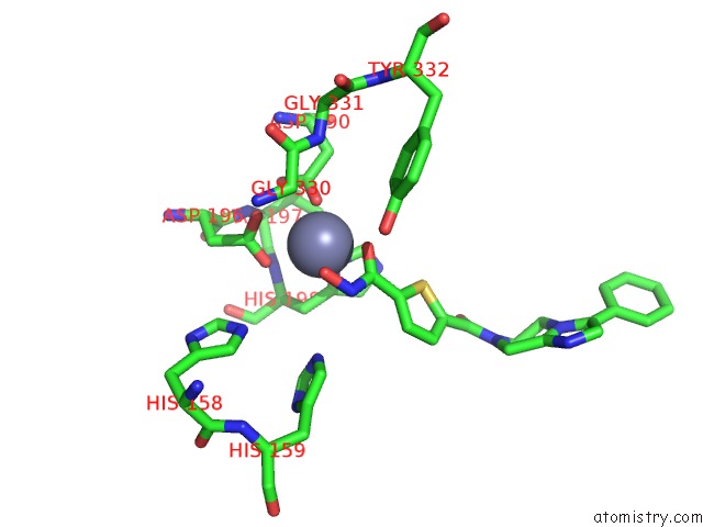
Mono view
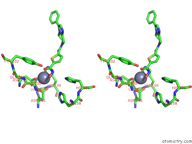
Stereo pair view
Mono view
Stereo pair view
A full contact list of Zinc with other atoms in the Zn binding
site number 1 of Structure of HDAC4 Catalytic Domain with A Gain-of-Function Mutation Bound to A Hydroxamic Acid Inhibitor within 5.0Å range:
|
Zinc binding site 2 out of 2 in 2vqv
Go back to
Zinc binding site 2 out
of 2 in the Structure of HDAC4 Catalytic Domain with A Gain-of-Function Mutation Bound to A Hydroxamic Acid Inhibitor
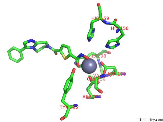
Mono view
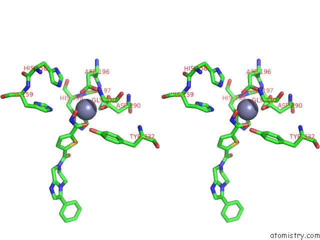
Stereo pair view
Mono view
Stereo pair view
A full contact list of Zinc with other atoms in the Zn binding
site number 2 of Structure of HDAC4 Catalytic Domain with A Gain-of-Function Mutation Bound to A Hydroxamic Acid Inhibitor within 5.0Å range:
|
Reference:
M.J.Bottomley,
P.Lo Surdo,
P.Di Giovine,
A.Cirillo,
R.Scarpelli,
F.Ferrigno,
P.Jones,
P.Neddermann,
R.De Francesco,
C.Steinkuhler,
P.Gallinari,
A.Carfi.
Structural and Functional Analysis of the Human HDAC4 Catalytic Domain Reveals A Regulatory Structural Zinc-Binding Domain. J.Biol.Chem. V. 283 26694 2008.
ISSN: ISSN 0021-9258
PubMed: 18614528
DOI: 10.1074/JBC.M803514200
Page generated: Thu Oct 17 04:28:13 2024
ISSN: ISSN 0021-9258
PubMed: 18614528
DOI: 10.1074/JBC.M803514200
Last articles
Zn in 9J0NZn in 9J0O
Zn in 9J0P
Zn in 9FJX
Zn in 9EKB
Zn in 9C0F
Zn in 9CAH
Zn in 9CH0
Zn in 9CH3
Zn in 9CH1