Zinc »
PDB 2vhf-2vr2 »
2vjz »
Zinc in PDB 2vjz: Crystal Structure Form Ultalente Insulin Microcrystals
Protein crystallography data
The structure of Crystal Structure Form Ultalente Insulin Microcrystals, PDB code: 2vjz
was solved by
A.Wagner,
J.Diez,
C.Schulze-Briese,
G.Schluckebier,
with X-Ray Crystallography technique. A brief refinement statistics is given in the table below:
| Resolution Low / High (Å) | 32.43 / 1.80 |
| Space group | H 3 |
| Cell size a, b, c (Å), α, β, γ (°) | 79.820, 79.820, 36.710, 90.00, 90.00, 120.00 |
| R / Rfree (%) | 17.2 / 23.9 |
Other elements in 2vjz:
The structure of Crystal Structure Form Ultalente Insulin Microcrystals also contains other interesting chemical elements:
| Chlorine | (Cl) | 4 atoms |
Zinc Binding Sites:
The binding sites of Zinc atom in the Crystal Structure Form Ultalente Insulin Microcrystals
(pdb code 2vjz). This binding sites where shown within
5.0 Angstroms radius around Zinc atom.
In total 2 binding sites of Zinc where determined in the Crystal Structure Form Ultalente Insulin Microcrystals, PDB code: 2vjz:
Jump to Zinc binding site number: 1; 2;
In total 2 binding sites of Zinc where determined in the Crystal Structure Form Ultalente Insulin Microcrystals, PDB code: 2vjz:
Jump to Zinc binding site number: 1; 2;
Zinc binding site 1 out of 2 in 2vjz
Go back to
Zinc binding site 1 out
of 2 in the Crystal Structure Form Ultalente Insulin Microcrystals
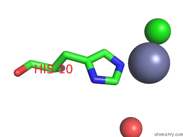
Mono view
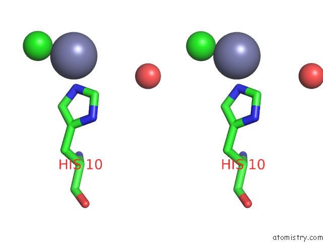
Stereo pair view
Mono view
Stereo pair view
A full contact list of Zinc with other atoms in the Zn binding
site number 1 of Crystal Structure Form Ultalente Insulin Microcrystals within 5.0Å range:
|
Zinc binding site 2 out of 2 in 2vjz
Go back to
Zinc binding site 2 out
of 2 in the Crystal Structure Form Ultalente Insulin Microcrystals
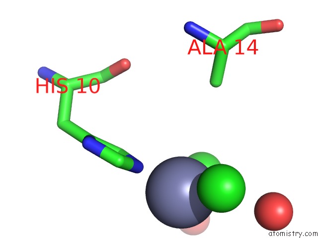
Mono view
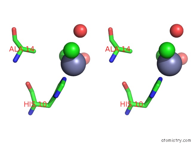
Stereo pair view
Mono view
Stereo pair view
A full contact list of Zinc with other atoms in the Zn binding
site number 2 of Crystal Structure Form Ultalente Insulin Microcrystals within 5.0Å range:
|
Reference:
A.Wagner,
J.Diez,
C.Schulze-Briese,
G.Schluckebier.
Crystal Structure of Ultralente-A Microcrystalline Insulin Suspension. Proteins V. 74 1018 2009.
ISSN: ISSN 0887-3585
PubMed: 18767151
DOI: 10.1002/PROT.22213
Page generated: Thu Oct 17 04:19:52 2024
ISSN: ISSN 0887-3585
PubMed: 18767151
DOI: 10.1002/PROT.22213
Last articles
Zn in 9J0NZn in 9J0O
Zn in 9J0P
Zn in 9FJX
Zn in 9EKB
Zn in 9C0F
Zn in 9CAH
Zn in 9CH0
Zn in 9CH3
Zn in 9CH1