Zinc »
PDB 2r1w-2rgv »
2rgv »
Zinc in PDB 2rgv: The Crystal Structure of Perr-Ox Highlights 2-Oxo-Histidine Formation
Protein crystallography data
The structure of The Crystal Structure of Perr-Ox Highlights 2-Oxo-Histidine Formation, PDB code: 2rgv
was solved by
D.A.K.Traore,
with X-Ray Crystallography technique. A brief refinement statistics is given in the table below:
| Resolution Low / High (Å) | 36.00 / 2.05 |
| Space group | P 1 |
| Cell size a, b, c (Å), α, β, γ (°) | 41.250, 42.700, 53.020, 82.05, 79.98, 61.63 |
| R / Rfree (%) | 21.1 / 30.8 |
Zinc Binding Sites:
The binding sites of Zinc atom in the The Crystal Structure of Perr-Ox Highlights 2-Oxo-Histidine Formation
(pdb code 2rgv). This binding sites where shown within
5.0 Angstroms radius around Zinc atom.
In total 2 binding sites of Zinc where determined in the The Crystal Structure of Perr-Ox Highlights 2-Oxo-Histidine Formation, PDB code: 2rgv:
Jump to Zinc binding site number: 1; 2;
In total 2 binding sites of Zinc where determined in the The Crystal Structure of Perr-Ox Highlights 2-Oxo-Histidine Formation, PDB code: 2rgv:
Jump to Zinc binding site number: 1; 2;
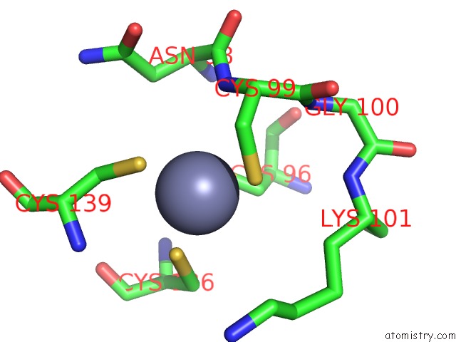
Zinc binding site 1 out of 2 in 2rgv
Go back to
Zinc binding site 1 out
of 2 in the The Crystal Structure of Perr-Ox Highlights 2-Oxo-Histidine Formation

Mono view

Stereo pair view
Mono view
Stereo pair view
A full contact list of Zinc with other atoms in the Zn binding
site number 1 of The Crystal Structure of Perr-Ox Highlights 2-Oxo-Histidine Formation within 5.0Å range:
|
Zinc binding site 2 out of 2 in 2rgv
Go back to
Zinc binding site 2 out
of 2 in the The Crystal Structure of Perr-Ox Highlights 2-Oxo-Histidine Formation
Mono view

Stereo pair view
Mono view
Stereo pair view
A full contact list of Zinc with other atoms in the Zn binding
site number 2 of The Crystal Structure of Perr-Ox Highlights 2-Oxo-Histidine Formation within 5.0Å range:
|
Reference:
D.A.Traore,
A.El Ghazouani,
L.Jacquamet,
F.Borel,
J.L.Ferrer,
D.Lascoux,
J.L.Ravanat,
M.Jaquinod,
G.Blondin,
C.Caux-Thang,
V.Duarte,
J.M.Latour.
Structural and Functional Characterization of 2-Oxo-Histidine in Oxidized Perr Protein. Nat.Chem.Biol. V. 5 53 2009.
ISSN: ISSN 1552-4450
PubMed: 19079268
DOI: 10.1038/NCHEMBIO.133
Page generated: Wed Aug 20 05:38:14 2025
ISSN: ISSN 1552-4450
PubMed: 19079268
DOI: 10.1038/NCHEMBIO.133
Last articles
Zn in 3KYAZn in 3KYD
Zn in 3KYC
Zn in 3KY9
Zn in 3KWC
Zn in 3KRY
Zn in 3KWE
Zn in 3KWD
Zn in 3KWA
Zn in 3KVT