Zinc »
PDB 2qzr-2rgt »
2r59 »
Zinc in PDB 2r59: Leukotriene A4 Hydrolase Complexed with Inhibitor RB3041
Enzymatic activity of Leukotriene A4 Hydrolase Complexed with Inhibitor RB3041
All present enzymatic activity of Leukotriene A4 Hydrolase Complexed with Inhibitor RB3041:
3.3.2.6;
3.3.2.6;
Protein crystallography data
The structure of Leukotriene A4 Hydrolase Complexed with Inhibitor RB3041, PDB code: 2r59
was solved by
F.Tholander,
J.Z.Haeggstrom,
M.Thunnissen,
A.Muroya,
B.P.Roques,
M.C.Fournie-Zaluski,
with X-Ray Crystallography technique. A brief refinement statistics is given in the table below:
| Resolution Low / High (Å) | 14.85 / 1.89 |
| Space group | P 21 21 21 |
| Cell size a, b, c (Å), α, β, γ (°) | 78.210, 87.230, 99.230, 90.00, 90.00, 90.00 |
| R / Rfree (%) | 17.5 / 22.7 |
Other elements in 2r59:
The structure of Leukotriene A4 Hydrolase Complexed with Inhibitor RB3041 also contains other interesting chemical elements:
| Ytterbium | (Yb) | 1 atom |
Zinc Binding Sites:
The binding sites of Zinc atom in the Leukotriene A4 Hydrolase Complexed with Inhibitor RB3041
(pdb code 2r59). This binding sites where shown within
5.0 Angstroms radius around Zinc atom.
In total only one binding site of Zinc was determined in the Leukotriene A4 Hydrolase Complexed with Inhibitor RB3041, PDB code: 2r59:
In total only one binding site of Zinc was determined in the Leukotriene A4 Hydrolase Complexed with Inhibitor RB3041, PDB code: 2r59:
Zinc binding site 1 out of 1 in 2r59
Go back to
Zinc binding site 1 out
of 1 in the Leukotriene A4 Hydrolase Complexed with Inhibitor RB3041
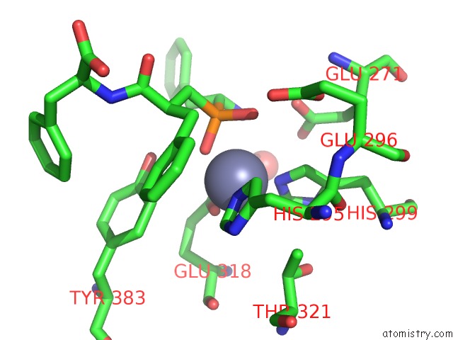
Mono view
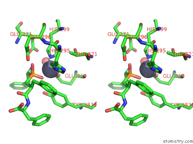
Stereo pair view
Mono view
Stereo pair view
A full contact list of Zinc with other atoms in the Zn binding
site number 1 of Leukotriene A4 Hydrolase Complexed with Inhibitor RB3041 within 5.0Å range:
|
Reference:
F.Tholander,
A.Muroya,
B.P.Roques,
M.C.Fournie-Zaluski,
M.M.Thunnissen,
J.Z.Haeggstrom.
Structure-Based Dissection of the Active Site Chemistry of Leukotriene A4 Hydrolase: Implications For M1 Aminopeptidases and Inhibitor Design. Chem.Biol. V. 15 920 2008.
ISSN: ISSN 1074-5521
PubMed: 18804029
DOI: 10.1016/J.CHEMBIOL.2008.07.018
Page generated: Thu Oct 17 03:39:50 2024
ISSN: ISSN 1074-5521
PubMed: 18804029
DOI: 10.1016/J.CHEMBIOL.2008.07.018
Last articles
Zn in 9J0NZn in 9J0O
Zn in 9J0P
Zn in 9FJX
Zn in 9EKB
Zn in 9C0F
Zn in 9CAH
Zn in 9CH0
Zn in 9CH3
Zn in 9CH1