Zinc in PDB 2q1z: Crystal Structure of Rhodobacter Sphaeroides Sige in Complex with the Anti-Sigma Chrr
Protein crystallography data
The structure of Crystal Structure of Rhodobacter Sphaeroides Sige in Complex with the Anti-Sigma Chrr, PDB code: 2q1z
was solved by
E.A.Campbell,
S.A.Darst,
with X-Ray Crystallography technique. A brief refinement statistics is given in the table below:
| Resolution Low / High (Å) | 35.00 / 2.40 |
| Space group | I 2 2 2 |
| Cell size a, b, c (Å), α, β, γ (°) | 43.601, 119.640, 280.659, 90.00, 90.00, 90.00 |
| R / Rfree (%) | 28.4 / 32.9 |
Zinc Binding Sites:
The binding sites of Zinc atom in the Crystal Structure of Rhodobacter Sphaeroides Sige in Complex with the Anti-Sigma Chrr
(pdb code 2q1z). This binding sites where shown within
5.0 Angstroms radius around Zinc atom.
In total 4 binding sites of Zinc where determined in the Crystal Structure of Rhodobacter Sphaeroides Sige in Complex with the Anti-Sigma Chrr, PDB code: 2q1z:
Jump to Zinc binding site number: 1; 2; 3; 4;
In total 4 binding sites of Zinc where determined in the Crystal Structure of Rhodobacter Sphaeroides Sige in Complex with the Anti-Sigma Chrr, PDB code: 2q1z:
Jump to Zinc binding site number: 1; 2; 3; 4;
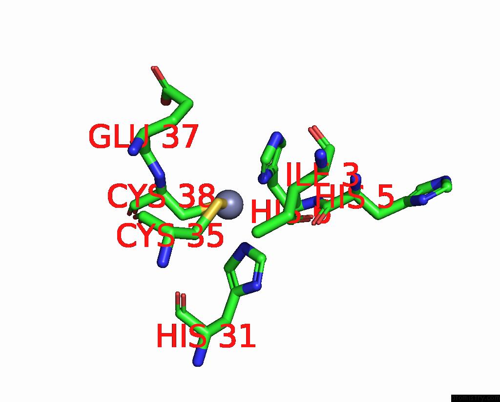
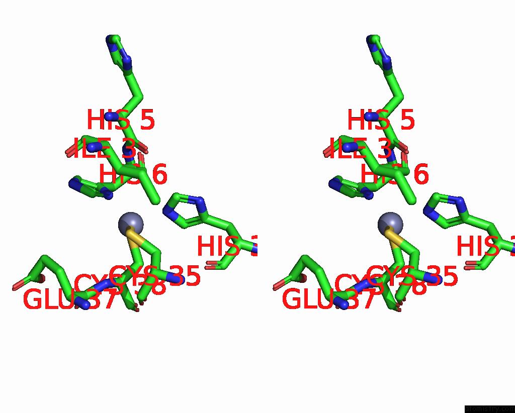
Zinc binding site 1 out of 4 in 2q1z
Go back to
Zinc binding site 1 out
of 4 in the Crystal Structure of Rhodobacter Sphaeroides Sige in Complex with the Anti-Sigma Chrr
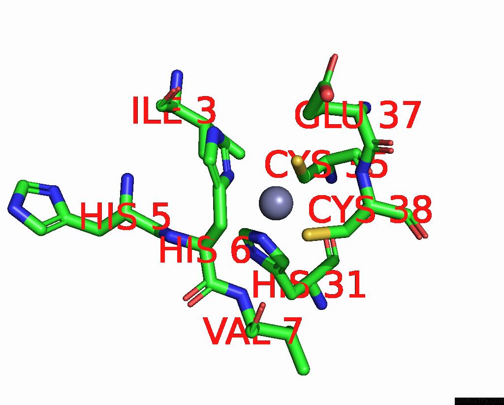
Mono view
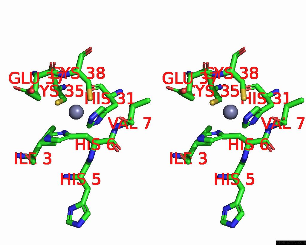
Stereo pair view
Mono view
Stereo pair view
A full contact list of Zinc with other atoms in the Zn binding
site number 1 of Crystal Structure of Rhodobacter Sphaeroides Sige in Complex with the Anti-Sigma Chrr within 5.0Å range:
|
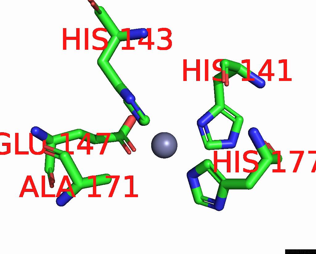
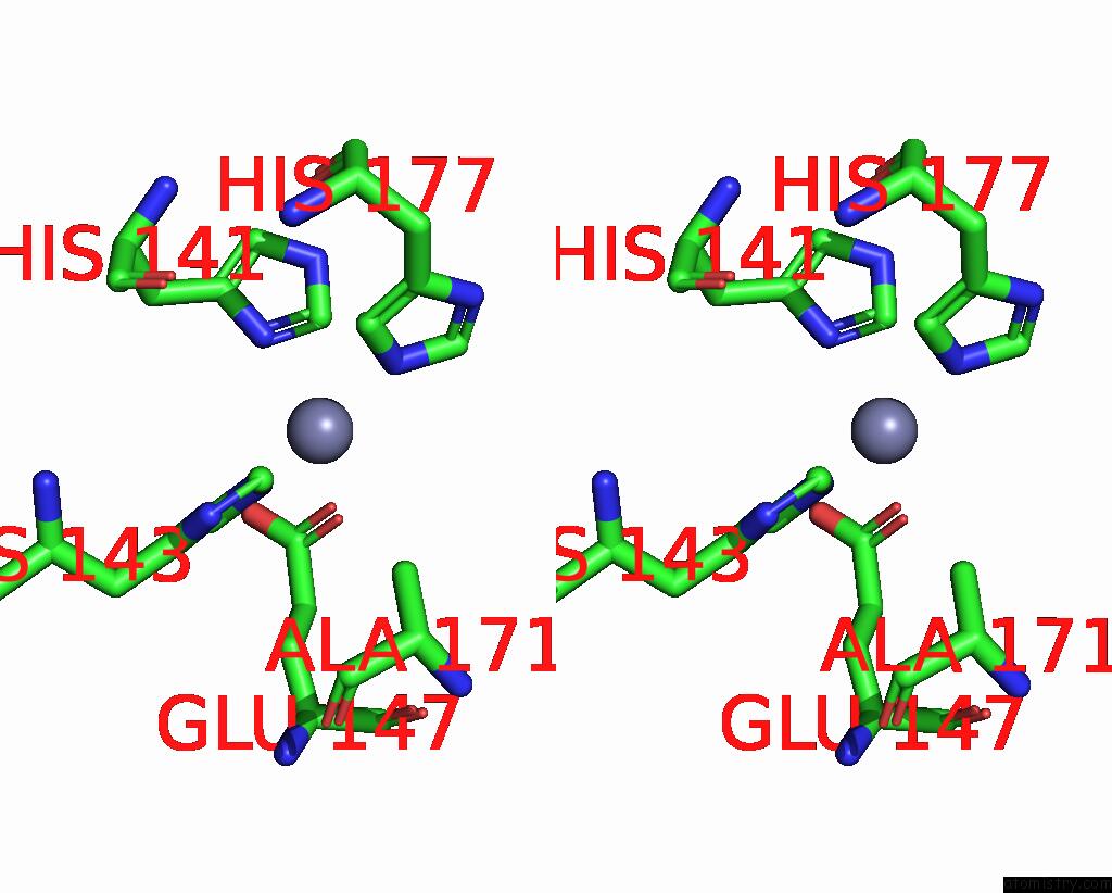
Zinc binding site 2 out of 4 in 2q1z
Go back to
Zinc binding site 2 out
of 4 in the Crystal Structure of Rhodobacter Sphaeroides Sige in Complex with the Anti-Sigma Chrr
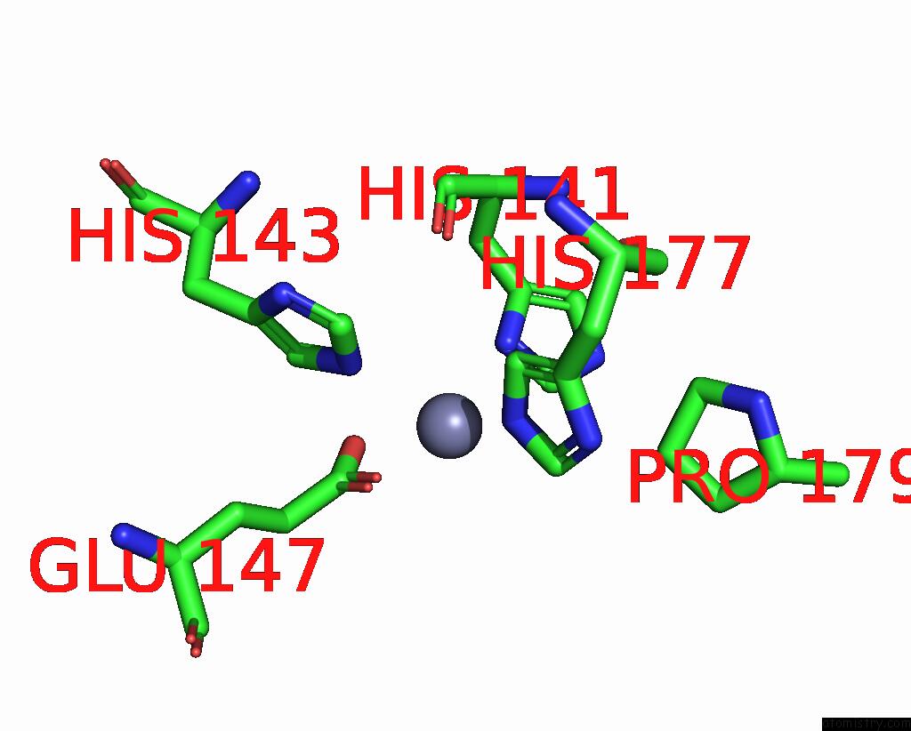
Mono view
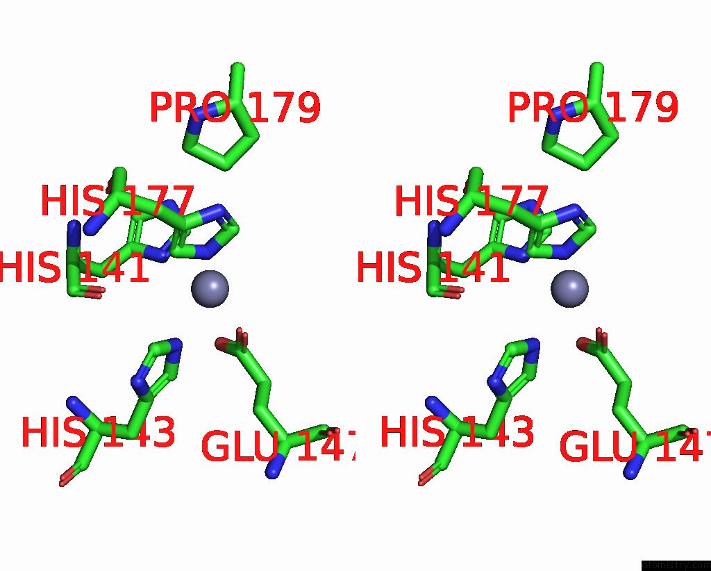
Stereo pair view
Mono view
Stereo pair view
A full contact list of Zinc with other atoms in the Zn binding
site number 2 of Crystal Structure of Rhodobacter Sphaeroides Sige in Complex with the Anti-Sigma Chrr within 5.0Å range:
|
Zinc binding site 3 out of 4 in 2q1z
Go back to
Zinc binding site 3 out
of 4 in the Crystal Structure of Rhodobacter Sphaeroides Sige in Complex with the Anti-Sigma Chrr
Mono view
Stereo pair view
Mono view
Stereo pair view
A full contact list of Zinc with other atoms in the Zn binding
site number 3 of Crystal Structure of Rhodobacter Sphaeroides Sige in Complex with the Anti-Sigma Chrr within 5.0Å range:
|
Zinc binding site 4 out of 4 in 2q1z
Go back to
Zinc binding site 4 out
of 4 in the Crystal Structure of Rhodobacter Sphaeroides Sige in Complex with the Anti-Sigma Chrr
Mono view
Stereo pair view
Mono view
Stereo pair view
A full contact list of Zinc with other atoms in the Zn binding
site number 4 of Crystal Structure of Rhodobacter Sphaeroides Sige in Complex with the Anti-Sigma Chrr within 5.0Å range:
|
Reference:
E.A.Campbell,
R.Greenwell,
J.R.Anthony,
S.Wang,
L.Lim,
K.Das,
H.J.Sofia,
T.J.Donohue,
S.A.Darst.
A Conserved Structural Module Regulates Transcriptional Responses to Diverse Stress Signals in Bacteria. Mol.Cell V. 27 793 2007.
ISSN: ISSN 1097-2765
PubMed: 17803943
DOI: 10.1016/J.MOLCEL.2007.07.009
Page generated: Thu Oct 17 03:20:52 2024
ISSN: ISSN 1097-2765
PubMed: 17803943
DOI: 10.1016/J.MOLCEL.2007.07.009
Last articles
Zn in 9MJ5Zn in 9HNW
Zn in 9G0L
Zn in 9FNE
Zn in 9DZN
Zn in 9E0I
Zn in 9D32
Zn in 9DAK
Zn in 8ZXC
Zn in 8ZUF