Zinc »
PDB 2o8h-2omh »
2ofi »
Zinc in PDB 2ofi: Crystal Structure of 3-Methyladenine Dna Glycosylase I (Tag) Bound to Dna/3MA
Protein crystallography data
The structure of Crystal Structure of 3-Methyladenine Dna Glycosylase I (Tag) Bound to Dna/3MA, PDB code: 2ofi
was solved by
A.H.Metz,
T.Hollis,
B.F.Eichman,
with X-Ray Crystallography technique. A brief refinement statistics is given in the table below:
| Resolution Low / High (Å) | 50.00 / 1.85 |
| Space group | P 63 |
| Cell size a, b, c (Å), α, β, γ (°) | 101.963, 101.963, 55.461, 90.00, 90.00, 120.00 |
| R / Rfree (%) | 17.6 / 19.8 |
Other elements in 2ofi:
The structure of Crystal Structure of 3-Methyladenine Dna Glycosylase I (Tag) Bound to Dna/3MA also contains other interesting chemical elements:
| Sodium | (Na) | 1 atom |
Zinc Binding Sites:
The binding sites of Zinc atom in the Crystal Structure of 3-Methyladenine Dna Glycosylase I (Tag) Bound to Dna/3MA
(pdb code 2ofi). This binding sites where shown within
5.0 Angstroms radius around Zinc atom.
In total only one binding site of Zinc was determined in the Crystal Structure of 3-Methyladenine Dna Glycosylase I (Tag) Bound to Dna/3MA, PDB code: 2ofi:
In total only one binding site of Zinc was determined in the Crystal Structure of 3-Methyladenine Dna Glycosylase I (Tag) Bound to Dna/3MA, PDB code: 2ofi:
Zinc binding site 1 out of 1 in 2ofi
Go back to
Zinc binding site 1 out
of 1 in the Crystal Structure of 3-Methyladenine Dna Glycosylase I (Tag) Bound to Dna/3MA
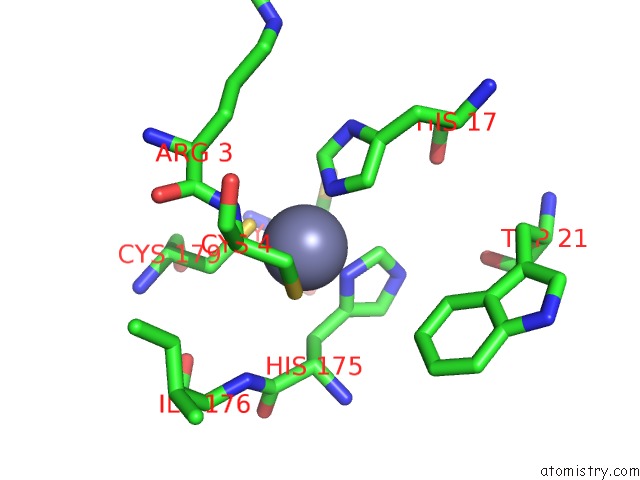
Mono view
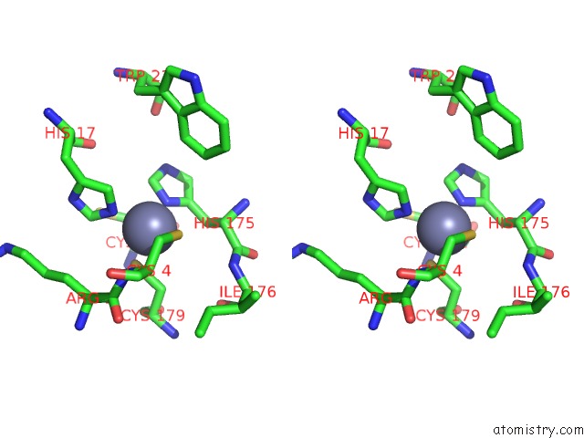
Stereo pair view
Mono view
Stereo pair view
A full contact list of Zinc with other atoms in the Zn binding
site number 1 of Crystal Structure of 3-Methyladenine Dna Glycosylase I (Tag) Bound to Dna/3MA within 5.0Å range:
|
Reference:
A.H.Metz,
T.Hollis,
B.F.Eichman.
Dna Damage Recognition and Repair By 3-Methyladenine Dna Glycosylase I (Tag). Embo J. V. 26 2411 2007.
ISSN: ISSN 0261-4189
PubMed: 17410210
DOI: 10.1038/SJ.EMBOJ.7601649
Page generated: Thu Oct 17 02:37:34 2024
ISSN: ISSN 0261-4189
PubMed: 17410210
DOI: 10.1038/SJ.EMBOJ.7601649
Last articles
Zn in 9J0NZn in 9J0O
Zn in 9J0P
Zn in 9FJX
Zn in 9EKB
Zn in 9C0F
Zn in 9CAH
Zn in 9CH0
Zn in 9CH3
Zn in 9CH1