Zinc »
PDB 2o8h-2omh »
2oc2 »
Zinc in PDB 2oc2: Structure of Testis Ace with RXPA380
Enzymatic activity of Structure of Testis Ace with RXPA380
All present enzymatic activity of Structure of Testis Ace with RXPA380:
3.4.15.1;
3.4.15.1;
Protein crystallography data
The structure of Structure of Testis Ace with RXPA380, PDB code: 2oc2
was solved by
H.R.Corradi,
C.S.Anthony,
S.L.Schwager,
P.Redelinghuys,
D.Georgiadis,
V.Dive,
K.R.Acharya,
E.D.Sturrock,
with X-Ray Crystallography technique. A brief refinement statistics is given in the table below:
| Resolution Low / High (Å) | 17.12 / 2.25 |
| Space group | P 21 21 21 |
| Cell size a, b, c (Å), α, β, γ (°) | 56.514, 84.763, 133.483, 90.00, 90.00, 90.00 |
| R / Rfree (%) | 21.5 / 26.2 |
Other elements in 2oc2:
The structure of Structure of Testis Ace with RXPA380 also contains other interesting chemical elements:
| Chlorine | (Cl) | 2 atoms |
Zinc Binding Sites:
The binding sites of Zinc atom in the Structure of Testis Ace with RXPA380
(pdb code 2oc2). This binding sites where shown within
5.0 Angstroms radius around Zinc atom.
In total only one binding site of Zinc was determined in the Structure of Testis Ace with RXPA380, PDB code: 2oc2:
In total only one binding site of Zinc was determined in the Structure of Testis Ace with RXPA380, PDB code: 2oc2:
Zinc binding site 1 out of 1 in 2oc2
Go back to
Zinc binding site 1 out
of 1 in the Structure of Testis Ace with RXPA380
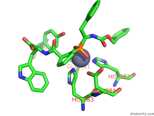
Mono view
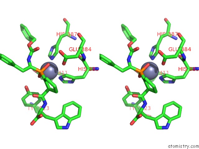
Stereo pair view
Mono view
Stereo pair view
A full contact list of Zinc with other atoms in the Zn binding
site number 1 of Structure of Testis Ace with RXPA380 within 5.0Å range:
|
Reference:
C.S.Anthony,
H.R.Corradi,
S.L.Schwager,
P.Redelinghuys,
D.Georgiadis,
V.Dive,
K.R.Acharya,
E.D.Sturrock.
The N Domain of Human Angiotensin-I Converting Enzyme: the Role of N-Glycosylation and the Crystal Structure in Complex with An N Domain Specific Phosphinic Inhibitor RXP407. J.Biol.Chem. V. 46 5473 2010.
ISSN: ISSN 0021-9258
PubMed: 20826823
DOI: 10.1074/JBC.M110.167866
Page generated: Thu Oct 17 02:35:14 2024
ISSN: ISSN 0021-9258
PubMed: 20826823
DOI: 10.1074/JBC.M110.167866
Last articles
Zn in 9J0NZn in 9J0O
Zn in 9J0P
Zn in 9FJX
Zn in 9EKB
Zn in 9C0F
Zn in 9CAH
Zn in 9CH0
Zn in 9CH3
Zn in 9CH1