Zinc »
PDB 2mmi-2n9p »
2n8a »
Zinc in PDB 2n8a: 1H, 13C and 15N Chemical Shift Assignments and Solution Structure For Parp-1 F1F2 Domains in Complex with A Dna Single-Strand Break
Enzymatic activity of 1H, 13C and 15N Chemical Shift Assignments and Solution Structure For Parp-1 F1F2 Domains in Complex with A Dna Single-Strand Break
All present enzymatic activity of 1H, 13C and 15N Chemical Shift Assignments and Solution Structure For Parp-1 F1F2 Domains in Complex with A Dna Single-Strand Break:
2.4.2.30;
2.4.2.30;
Zinc Binding Sites:
The binding sites of Zinc atom in the 1H, 13C and 15N Chemical Shift Assignments and Solution Structure For Parp-1 F1F2 Domains in Complex with A Dna Single-Strand Break
(pdb code 2n8a). This binding sites where shown within
5.0 Angstroms radius around Zinc atom.
In total 2 binding sites of Zinc where determined in the 1H, 13C and 15N Chemical Shift Assignments and Solution Structure For Parp-1 F1F2 Domains in Complex with A Dna Single-Strand Break, PDB code: 2n8a:
Jump to Zinc binding site number: 1; 2;
In total 2 binding sites of Zinc where determined in the 1H, 13C and 15N Chemical Shift Assignments and Solution Structure For Parp-1 F1F2 Domains in Complex with A Dna Single-Strand Break, PDB code: 2n8a:
Jump to Zinc binding site number: 1; 2;
Zinc binding site 1 out of 2 in 2n8a
Go back to
Zinc binding site 1 out
of 2 in the 1H, 13C and 15N Chemical Shift Assignments and Solution Structure For Parp-1 F1F2 Domains in Complex with A Dna Single-Strand Break

Mono view
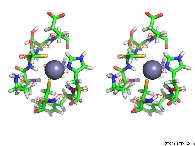
Stereo pair view
Mono view
Stereo pair view
A full contact list of Zinc with other atoms in the Zn binding
site number 1 of 1H, 13C and 15N Chemical Shift Assignments and Solution Structure For Parp-1 F1F2 Domains in Complex with A Dna Single-Strand Break within 5.0Å range:
|
Zinc binding site 2 out of 2 in 2n8a
Go back to
Zinc binding site 2 out
of 2 in the 1H, 13C and 15N Chemical Shift Assignments and Solution Structure For Parp-1 F1F2 Domains in Complex with A Dna Single-Strand Break
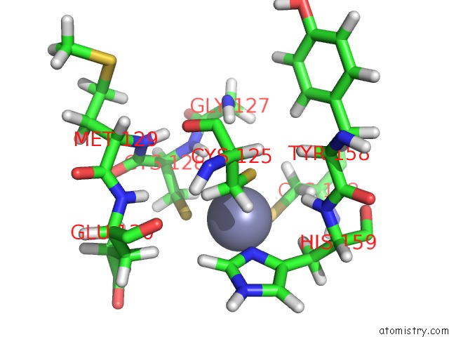
Mono view
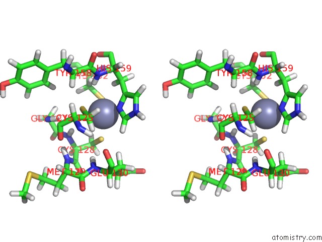
Stereo pair view
Mono view
Stereo pair view
A full contact list of Zinc with other atoms in the Zn binding
site number 2 of 1H, 13C and 15N Chemical Shift Assignments and Solution Structure For Parp-1 F1F2 Domains in Complex with A Dna Single-Strand Break within 5.0Å range:
|
Reference:
S.Eustermann,
W.F.Wu,
M.F.Langelier,
J.C.Yang,
L.E.Easton,
A.A.Riccio,
J.M.Pascal,
D.Neuhaus.
Structural Basis of Detection and Signaling of Dna Single-Strand Breaks By Human Parp-1. Mol.Cell V. 60 742 2015.
ISSN: ISSN 1097-2765
PubMed: 26626479
DOI: 10.1016/J.MOLCEL.2015.10.032
Page generated: Thu Oct 17 02:12:28 2024
ISSN: ISSN 1097-2765
PubMed: 26626479
DOI: 10.1016/J.MOLCEL.2015.10.032
Last articles
Zn in 9J0NZn in 9J0O
Zn in 9J0P
Zn in 9FJX
Zn in 9EKB
Zn in 9C0F
Zn in 9CAH
Zn in 9CH0
Zn in 9CH3
Zn in 9CH1