Zinc »
PDB 2jb0-2jtn »
2jg6 »
Zinc in PDB 2jg6: Crystal Structure of A 3-Methyladenine Dna Glycosylase I From Staphylococcus Aureus
Enzymatic activity of Crystal Structure of A 3-Methyladenine Dna Glycosylase I From Staphylococcus Aureus
All present enzymatic activity of Crystal Structure of A 3-Methyladenine Dna Glycosylase I From Staphylococcus Aureus:
3.2.2.20;
3.2.2.20;
Protein crystallography data
The structure of Crystal Structure of A 3-Methyladenine Dna Glycosylase I From Staphylococcus Aureus, PDB code: 2jg6
was solved by
X.Yan,
L.G.Carter,
H.Liu,
M.Dorward,
S.A.Mcmahon,
K.A.Johnson,
M.Oke,
P.J.Coote,
J.H.Naismith,
with X-Ray Crystallography technique. A brief refinement statistics is given in the table below:
| Resolution Low / High (Å) | 29.81 / 1.70 |
| Space group | C 1 2 1 |
| Cell size a, b, c (Å), α, β, γ (°) | 107.460, 63.098, 38.470, 90.00, 109.24, 90.00 |
| R / Rfree (%) | 16.3 / 19.3 |
Zinc Binding Sites:
The binding sites of Zinc atom in the Crystal Structure of A 3-Methyladenine Dna Glycosylase I From Staphylococcus Aureus
(pdb code 2jg6). This binding sites where shown within
5.0 Angstroms radius around Zinc atom.
In total only one binding site of Zinc was determined in the Crystal Structure of A 3-Methyladenine Dna Glycosylase I From Staphylococcus Aureus, PDB code: 2jg6:
In total only one binding site of Zinc was determined in the Crystal Structure of A 3-Methyladenine Dna Glycosylase I From Staphylococcus Aureus, PDB code: 2jg6:
Zinc binding site 1 out of 1 in 2jg6
Go back to
Zinc binding site 1 out
of 1 in the Crystal Structure of A 3-Methyladenine Dna Glycosylase I From Staphylococcus Aureus
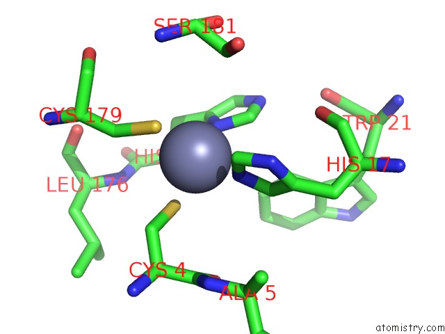
Mono view
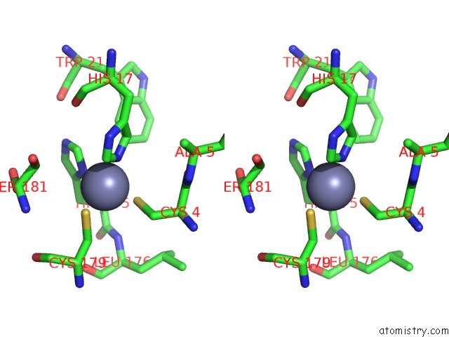
Stereo pair view
Mono view
Stereo pair view
A full contact list of Zinc with other atoms in the Zn binding
site number 1 of Crystal Structure of A 3-Methyladenine Dna Glycosylase I From Staphylococcus Aureus within 5.0Å range:
|
Reference:
M.Oke,
L.G.Carter,
K.A.Johnson,
H.Liu,
S.A.Mcmahon,
X.Yan,
M.Kerou,
N.D.Weikart,
N.Kadi,
M.A.Sheikh,
S.Schmelz,
M.Dorward,
M.Zawadzki,
C.Cozens,
H.Falconer,
H.Powers,
I.M.Overton,
C.A.J.Van Niekerk,
X.Peng,
P.Patel,
R.A.Garrett,
D.Prangishvili,
C.H.Botting,
P.J.Coote,
D.T.F.Dryden,
G.J.Barton,
U.Schwarz-Linek,
G.L.Challis,
G.L.Taylor,
M.F.White,
J.H.Naismith.
The Scottish Structural Proteomics Facility: Targets, Methods and Outputs. J.Struct.Funct.Genomics V. 11 167 2010.
ISSN: ISSN 1345-711X
PubMed: 20419351
DOI: 10.1007/S10969-010-9090-Y
Page generated: Wed Aug 20 03:47:25 2025
ISSN: ISSN 1345-711X
PubMed: 20419351
DOI: 10.1007/S10969-010-9090-Y
Last articles
Zn in 3DDGZn in 3DDF
Zn in 3DDB
Zn in 3DCP
Zn in 3DDA
Zn in 3DD8
Zn in 3DD0
Zn in 3DCW
Zn in 3DCS
Zn in 3DC8