Zinc »
PDB 2jb0-2jtn »
2jg4 »
Zinc in PDB 2jg4: Substrate-Free Ide Structure in Its Closed Conformation
Enzymatic activity of Substrate-Free Ide Structure in Its Closed Conformation
All present enzymatic activity of Substrate-Free Ide Structure in Its Closed Conformation:
3.4.24.56;
3.4.24.56;
Protein crystallography data
The structure of Substrate-Free Ide Structure in Its Closed Conformation, PDB code: 2jg4
was solved by
E.Malito,
W.J.Tang,
with X-Ray Crystallography technique. A brief refinement statistics is given in the table below:
| Resolution Low / High (Å) | 15.00 / 2.80 |
| Space group | P 65 |
| Cell size a, b, c (Å), α, β, γ (°) | 263.211, 263.211, 90.408, 90.00, 90.00, 120.00 |
| R / Rfree (%) | 18.2 / 22.7 |
Zinc Binding Sites:
The binding sites of Zinc atom in the Substrate-Free Ide Structure in Its Closed Conformation
(pdb code 2jg4). This binding sites where shown within
5.0 Angstroms radius around Zinc atom.
In total 2 binding sites of Zinc where determined in the Substrate-Free Ide Structure in Its Closed Conformation, PDB code: 2jg4:
Jump to Zinc binding site number: 1; 2;
In total 2 binding sites of Zinc where determined in the Substrate-Free Ide Structure in Its Closed Conformation, PDB code: 2jg4:
Jump to Zinc binding site number: 1; 2;
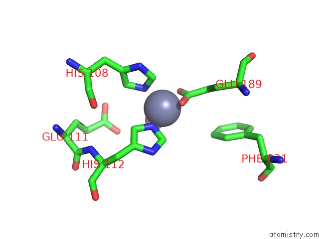
Zinc binding site 1 out of 2 in 2jg4
Go back to
Zinc binding site 1 out
of 2 in the Substrate-Free Ide Structure in Its Closed Conformation
Mono view

Stereo pair view
Mono view
Stereo pair view
A full contact list of Zinc with other atoms in the Zn binding
site number 1 of Substrate-Free Ide Structure in Its Closed Conformation within 5.0Å range:
|
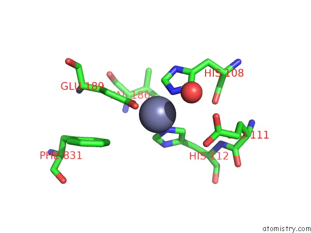
Zinc binding site 2 out of 2 in 2jg4
Go back to
Zinc binding site 2 out
of 2 in the Substrate-Free Ide Structure in Its Closed Conformation
Mono view
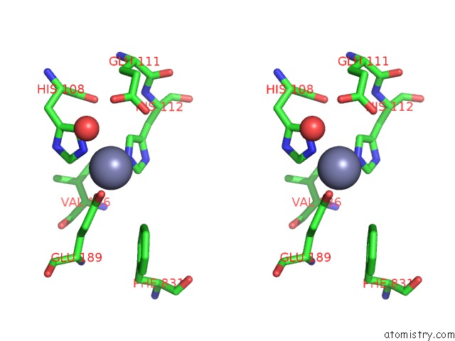
Stereo pair view
Mono view
Stereo pair view
A full contact list of Zinc with other atoms in the Zn binding
site number 2 of Substrate-Free Ide Structure in Its Closed Conformation within 5.0Å range:
|
Reference:
H.Im,
M.Manolopoulou,
E.Malito,
Y.Shen,
J.Zhao,
M.Neant-Fery,
C.-Y.Sun,
S.C.Meredith,
S.S.Sisodia,
M.A.Leissring,
W.J.Tang.
Structure of Substrate-Free Human Insulin Degrading Enzyme (Ide) and Biophysical Analysis of Atp-Induced Conformational Switch of Ide J.Biol.Chem. V. 282 25453 2007.
ISSN: ISSN 0021-9258
PubMed: 17613531
DOI: 10.1074/JBC.M701590200
Page generated: Wed Aug 20 03:47:26 2025
ISSN: ISSN 0021-9258
PubMed: 17613531
DOI: 10.1074/JBC.M701590200
Last articles
Zn in 3DDGZn in 3DDF
Zn in 3DDB
Zn in 3DCP
Zn in 3DDA
Zn in 3DD8
Zn in 3DD0
Zn in 3DCW
Zn in 3DCS
Zn in 3DC8