Zinc »
PDB 2ivh-2jaz »
2j13 »
Zinc in PDB 2j13: Structure of A Family 4 Carbohydrate Esterase From Bacillus Anthracis
Protein crystallography data
The structure of Structure of A Family 4 Carbohydrate Esterase From Bacillus Anthracis, PDB code: 2j13
was solved by
T.M.Gloster,
L.Oberbarnscheidt,
E.J.Taylor,
G.J.Davies,
with X-Ray Crystallography technique. A brief refinement statistics is given in the table below:
| Resolution Low / High (Å) | 30.00 / 1.7 |
| Space group | P 32 2 1 |
| Cell size a, b, c (Å), α, β, γ (°) | 56.927, 56.927, 158.531, 90.00, 90.00, 120.00 |
| R / Rfree (%) | 18.6 / 23 |
Other elements in 2j13:
The structure of Structure of A Family 4 Carbohydrate Esterase From Bacillus Anthracis also contains other interesting chemical elements:
| Arsenic | (As) | 1 atom |
Zinc Binding Sites:
The binding sites of Zinc atom in the Structure of A Family 4 Carbohydrate Esterase From Bacillus Anthracis
(pdb code 2j13). This binding sites where shown within
5.0 Angstroms radius around Zinc atom.
In total 4 binding sites of Zinc where determined in the Structure of A Family 4 Carbohydrate Esterase From Bacillus Anthracis, PDB code: 2j13:
Jump to Zinc binding site number: 1; 2; 3; 4;
In total 4 binding sites of Zinc where determined in the Structure of A Family 4 Carbohydrate Esterase From Bacillus Anthracis, PDB code: 2j13:
Jump to Zinc binding site number: 1; 2; 3; 4;
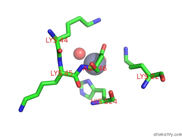
Zinc binding site 1 out of 4 in 2j13
Go back to
Zinc binding site 1 out
of 4 in the Structure of A Family 4 Carbohydrate Esterase From Bacillus Anthracis
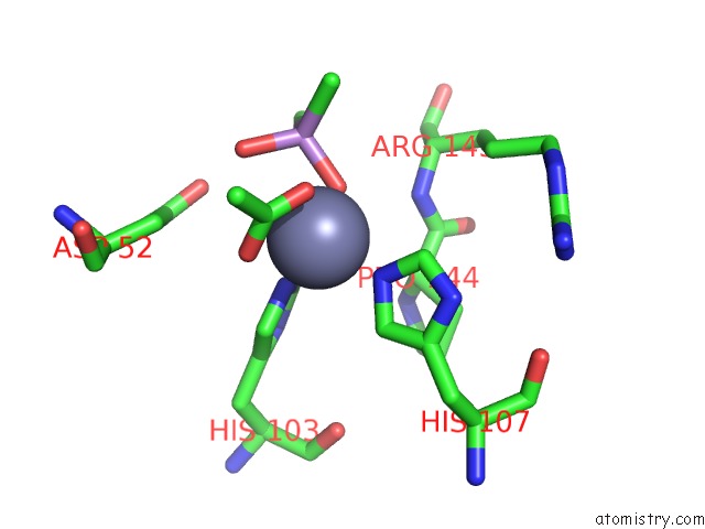
Mono view
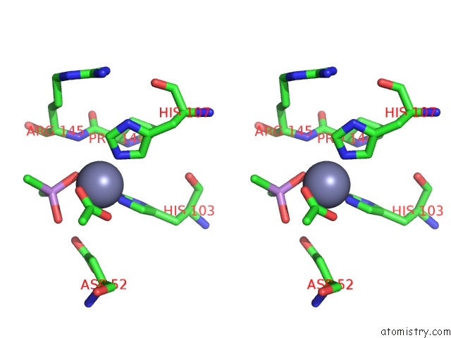
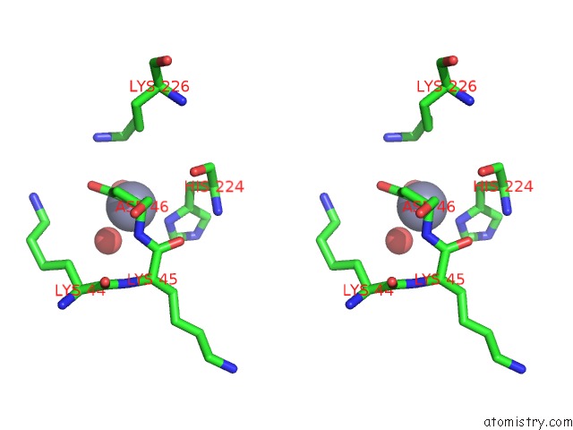
Stereo pair view
Mono view
Stereo pair view
A full contact list of Zinc with other atoms in the Zn binding
site number 1 of Structure of A Family 4 Carbohydrate Esterase From Bacillus Anthracis within 5.0Å range:
|
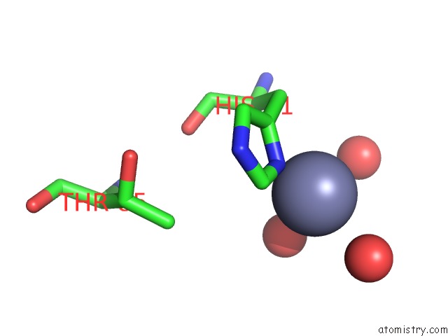
Zinc binding site 2 out of 4 in 2j13
Go back to
Zinc binding site 2 out
of 4 in the Structure of A Family 4 Carbohydrate Esterase From Bacillus Anthracis
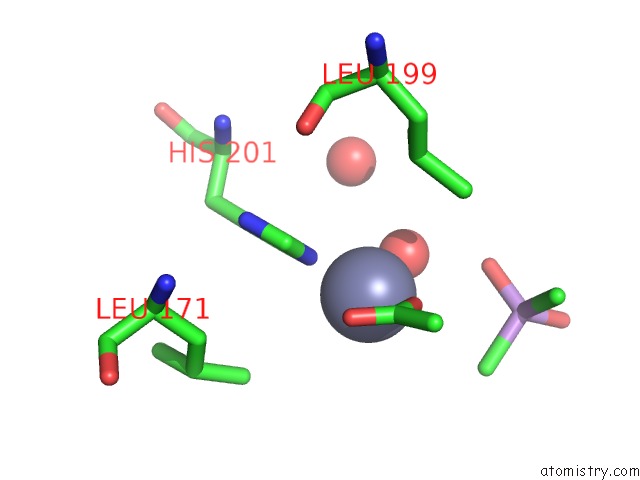
Mono view
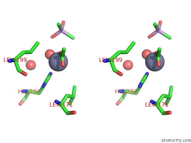
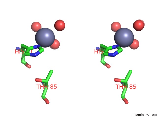
Stereo pair view
Mono view
Stereo pair view
A full contact list of Zinc with other atoms in the Zn binding
site number 2 of Structure of A Family 4 Carbohydrate Esterase From Bacillus Anthracis within 5.0Å range:
|
Zinc binding site 3 out of 4 in 2j13
Go back to
Zinc binding site 3 out
of 4 in the Structure of A Family 4 Carbohydrate Esterase From Bacillus Anthracis
Mono view
Stereo pair view
Mono view
Stereo pair view
A full contact list of Zinc with other atoms in the Zn binding
site number 3 of Structure of A Family 4 Carbohydrate Esterase From Bacillus Anthracis within 5.0Å range:
|
Zinc binding site 4 out of 4 in 2j13
Go back to
Zinc binding site 4 out
of 4 in the Structure of A Family 4 Carbohydrate Esterase From Bacillus Anthracis
Mono view
Stereo pair view
Mono view
Stereo pair view
A full contact list of Zinc with other atoms in the Zn binding
site number 4 of Structure of A Family 4 Carbohydrate Esterase From Bacillus Anthracis within 5.0Å range:
|
Reference:
L.Oberbarnscheidt,
E.J.Taylor,
G.J.Davies,
T.M.Gloster.
Structure of A Carbohydrate Esterase From Bacillus Anthracis. Proteins: Struct., Funct., V. 66 250 2007BIOINF..
ISSN: ISSN 0887-3585
PubMed: 17063474
DOI: 10.1002/PROT.21217
Page generated: Thu Oct 17 01:03:19 2024
ISSN: ISSN 0887-3585
PubMed: 17063474
DOI: 10.1002/PROT.21217
Last articles
Zn in 9J0NZn in 9J0O
Zn in 9J0P
Zn in 9FJX
Zn in 9EKB
Zn in 9C0F
Zn in 9CAH
Zn in 9CH0
Zn in 9CH3
Zn in 9CH1