Zinc »
PDB 2ics-2iv0 »
2isg »
Zinc in PDB 2isg: Botulinum Neurotoxin A Light Chain Wt Crystal Form B
Protein crystallography data
The structure of Botulinum Neurotoxin A Light Chain Wt Crystal Form B, PDB code: 2isg
was solved by
A.T.Brunger,
C.M.Stegmann,
with X-Ray Crystallography technique. A brief refinement statistics is given in the table below:
| Resolution Low / High (Å) | 42.44 / 2.00 |
| Space group | P 1 21 1 |
| Cell size a, b, c (Å), α, β, γ (°) | 41.991, 190.955, 56.949, 90.00, 90.09, 90.00 |
| R / Rfree (%) | 24 / 27.7 |
Other elements in 2isg:
The structure of Botulinum Neurotoxin A Light Chain Wt Crystal Form B also contains other interesting chemical elements:
| Nickel | (Ni) | 2 atoms |
Zinc Binding Sites:
The binding sites of Zinc atom in the Botulinum Neurotoxin A Light Chain Wt Crystal Form B
(pdb code 2isg). This binding sites where shown within
5.0 Angstroms radius around Zinc atom.
In total 2 binding sites of Zinc where determined in the Botulinum Neurotoxin A Light Chain Wt Crystal Form B, PDB code: 2isg:
Jump to Zinc binding site number: 1; 2;
In total 2 binding sites of Zinc where determined in the Botulinum Neurotoxin A Light Chain Wt Crystal Form B, PDB code: 2isg:
Jump to Zinc binding site number: 1; 2;
Zinc binding site 1 out of 2 in 2isg
Go back to
Zinc binding site 1 out
of 2 in the Botulinum Neurotoxin A Light Chain Wt Crystal Form B
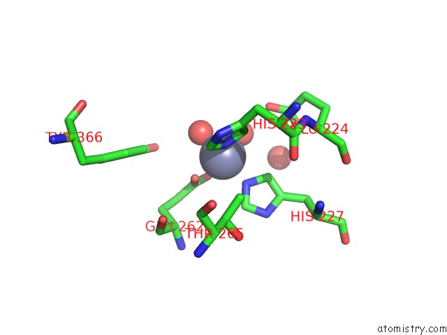
Mono view
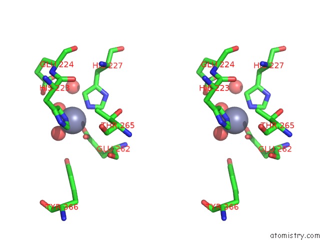
Stereo pair view
Mono view
Stereo pair view
A full contact list of Zinc with other atoms in the Zn binding
site number 1 of Botulinum Neurotoxin A Light Chain Wt Crystal Form B within 5.0Å range:
|
Zinc binding site 2 out of 2 in 2isg
Go back to
Zinc binding site 2 out
of 2 in the Botulinum Neurotoxin A Light Chain Wt Crystal Form B
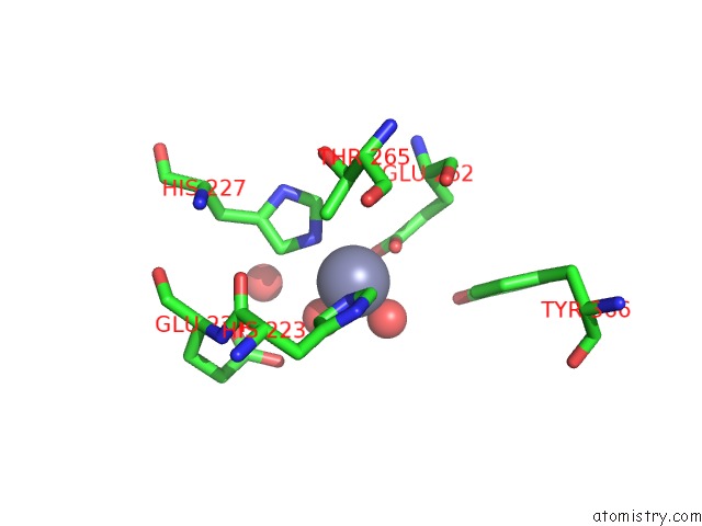
Mono view
Stereo pair view
Mono view
Stereo pair view
A full contact list of Zinc with other atoms in the Zn binding
site number 2 of Botulinum Neurotoxin A Light Chain Wt Crystal Form B within 5.0Å range:
|
Reference:
J.C.Burnett,
G.Ruthel,
C.M.Stegmann,
R.G.Panchal,
T.L.Nguyen,
A.R.Hermone,
R.G.Stafford,
D.J.Lane,
T.A.Kenny,
C.F.Mcgrath,
P.Wipf,
A.M.Stahl,
J.J.Schmidt,
R.Gussio,
A.T.Brunger,
S.Bavari.
Inhibition of Metalloprotease Botulinum Serotype A From A Pseudo-Peptide Binding Mode to A Small Molecule That Is Active in Primary Neurons. J.Biol.Chem. V. 282 5004 2007.
ISSN: ISSN 0021-9258
PubMed: 17092934
DOI: 10.1074/JBC.M608166200
Page generated: Thu Oct 17 00:59:00 2024
ISSN: ISSN 0021-9258
PubMed: 17092934
DOI: 10.1074/JBC.M608166200
Last articles
Zn in 9J0NZn in 9J0O
Zn in 9J0P
Zn in 9FJX
Zn in 9EKB
Zn in 9C0F
Zn in 9CAH
Zn in 9CH0
Zn in 9CH3
Zn in 9CH1